Image Analysis in Earth Sciences
Autor Renée Heilbronner, Steve Barretten Limba Engleză Hardback – 29 iul 2013
Evoluția geologiei moderne a fost marcată de trecerea de la observația calitativă la rigoarea analizei cantitative digitale. Observăm cum această transformare a impus necesitatea unor metodologii precise pentru interpretarea datelor vizuale complexe. Lucrarea Image Analysis in Earth Sciences, semnată de Renée Heilbronner și Steve Barrett, răspunde acestei nevoi, oferind un cadru tehnic structurat pentru cercetătorii care studiază microstructurile și texturile materialelor terestre. Volumul nu se limitează la teorie, ci integrează exemple practice lucrate, utilizând software-ul accesibil Image SXM pentru a ghida cititorul prin procese esențiale: de la achiziția imaginilor prin microscopie optică sau electronică, până la preprocesare și segmentarea obiectelor. Remarcăm atenția deosebită acordată analizei distribuției dimensiunilor granulelor și a fabricilor de suprafață, elemente critice în înțelegerea proceselor de deformare și cristalizare. Ultimele capitole extind discuția către analiza orientării cristalografice, reflectând cele mai noi standarde în petrologie. Cititorii familiarizați cu Quantitative Textural Measurements in Igneous and Metamorphic Petrology de Michael Denis Higgins vor aprecia modul în care acest volum aduce un plus de specificitate tehnică în zona procesării digitale a imaginilor, oferind soluții algoritmice acolo unde alte lucrări se rezumă la fundamentele petrologice. Recomandăm acest manual pentru structura sa modulară, care permite navigarea rapidă între conceptele de analiză în domeniul frecvenței (FFT) și aplicațiile practice de orientare a particulelor, fiind o resursă indispensabilă în laboratoarele de cercetare geologică.
Preț: 979.24 lei
Preț vechi: 1194.19 lei
-18%
Carte disponibilă
Livrare economică 15-29 august
Specificații
ISBN-10: 3642103421
Pagini: 544
Ilustrații: XIX, 520 p. 596 illus., 257 illus. in color.
Dimensiuni: 183 x 260 x 33 mm
Greutate: 1.33 kg
Ediția:2014
Editura: Springer
Locul publicării:Berlin, Heidelberg, Germany
V-ar putea interesa
-
 Snow CrystalsW. A. BentleyPreț: 160.69 lei
 -
 Sand Up Close: Aesthetics, Philosophy, and ScienceOliver LenzenPreț: 299.51 lei
 -
 Atlas of Minerals and Igneous and Metamorphic Rocks in Thin-SectionAlessandro Da MommioPreț: 441.31 lei
 -
 XAFS for EveryoneScott Calvin-9%Preț: 816.66 lei897.42 lei
 -
 Structural GeologyHaakon Fossen-8%Preț: 472.93 lei514.05 lei
 -
 Remote Sensing and Image InterpretationThomas Lillesand-18%Preț: 1345.99 lei1641.45 lei
 -
 Seismic Data Interpretation Using Digital Image ProcessingAbdullatif A Al-Shuhail-23%Preț: 608.18 lei789.84 lei
 -
 Microanalysis of Atmospheric ParticlesJoseph M. Conny-23%Preț: 960.36 lei1247.22 lei
 -
 Image ProcessingMaria M P Petrou-14%Preț: 807.82 lei939.33 lei
 -
 Rocks and Minerals in Thin Section: A Colour AtlasW.S. MacKenziePreț: 372.37 lei
 -
 Digital Image Processing, Global EditionRafael Gonzalez-11%Preț: 652.86 lei733.54 lei
 -
 MicrocosmosBrandon BrollPreț: 142.45 lei
 -
 Preț: 314.40 lei
 -
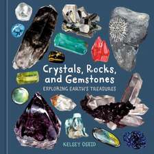 Crystals, Rocks, and GemstonesKelsey OseidPreț: 108.64 lei
 -
 Materials Data ScienceStefan Sandfeld-15%Preț: 573.37 lei674.56 lei
 -
 -18%Preț: 968.95 lei1181.65 lei
 -
 MATLAB® Recipes for Earth SciencesMartin H. Trauth-18%Preț: 737.69 lei899.63 lei
 -
 Computer Analysis of Images and PatternsModesto Castrillón-Santana-20%Preț: 327.69 lei409.62 lei
 -
 Intelligent Analysis of Optical ImagesZhengjun Liu-18%Preț: 867.54 lei1057.97 lei
Public țintă
ResearchDe ce să citești această carte
Recomandăm această lucrare cercetătorilor și studenților la nivel masteral sau doctoral care doresc să stăpânească instrumentele digitale de cuantificare a texturilor geologice. Cititorul câștigă o metodologie clară de utilizare a software-ului Image SXM, transformând observațiile microscopice în date statistice riguroase. Este motivul principal pentru care acest volum publicat de Springer rămâne un reper în petrologia modernă.
Despre autor
Renée Heilbronner este un expert recunoscut în analiza microstructurilor, activând în cadrul universităților din Basel și Tromsø, unde s-a specializat în mecanismele de deformare ale rocilor. Steve Barrett aduce expertiza tehnică necesară din perspectiva fizicii și a dezvoltării de software, fiind creatorul Image SXM, o variantă a ImageJ adaptată pentru microscopie. Colaborarea lor îmbină riguroarea științelor pământului cu precizia informatică, oferind un instrument complet pentru analiza materialelor naturale.
Descriere scurtă
Cuprins
Notă biografică
http://pages.unibas.ch/earth/micro
Dr Steve Barrett is the author of the internationally renowned image analysis software Image SXM. He has been developing the software continuously over the past two decades, from its origins as a spin-off from the freeware NIH Image, to the extensions that allow it to handle images from over fifty types of optical and scanning microscopes. A customized version of this software (PrinCIPia) based on the CIP method can handle the calculation, display, analysis and manipulation of images representing the crystallographic orientation of grains in rock samples imaged by polarizing microscopes. He has published widely in the field of nanoscience and has also collaborated with medics to create microscopy image analysis software for medical applications (MIASMA). He has over twenty years experience teaching to undergraduates and postgraduates. http://www.liv.ac.uk/~sdb
Caracteristici
Much needed book for earth and material scientists as well as university teaching programmes
Based on free software Image SXM
Includes supplementary material: sn.pub/extras