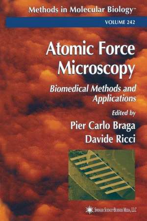
Atomic Force Microscopy: Biomedical Methods and Applications: Methods in Molecular Biology, cartea 242
Editat de Pier Carlo Braga, Davide Riccien Limba Engleză Paperback – 11 aug 2013
Din seria Methods in Molecular Biology
- 15%
 Preț: 629.45 lei
 - 15%
 Preț: 627.45 lei
 - 5%
 Preț: 1014.54 lei
 - 18%
 Preț: 2039.44 lei
 - 5%
 Preț: 1248.71 lei
 - 18%
 Preț: 1336.11 lei
 - 18%
 Preț: 1341.26 lei
 - 18%
 Preț: 1085.72 lei
 - 18%
 Preț: 1353.39 lei
 - 18%
 Preț: 1081.17 lei
 - 18%
 Preț: 1186.33 lei
 - 5%
 Preț: 1249.45 lei
 - 5%
 Preț: 1065.47 lei
 - 5%
 Preț: 788.57 lei
 - 5%
 Preț: 1576.70 lei
 - 5%
 Preț: 1073.37 lei
 - 5%
 Preț: 1361.54 lei
 - 18%
 Preț: 771.93 lei
 - 18%
 Preț: 749.09 lei
 - 18%
 Preț: 753.63 lei
 - 18%
 Preț: 1083.07 lei
 - 5%
 Preț: 1257.85 lei
 - 18%
 Preț: 1070.58 lei
 - 15%
 Preț: 669.59 lei
 - 18%
 Preț: 1076.62 lei
 - 18%
 Preț: 678.44 lei
 - 18%
 Preț: 678.44 lei
 - 18%
 Preț: 1343.97 lei
 - 5%
 Preț: 1556.10 lei
 - 5%
 Preț: 1554.98 lei
 - 5%
 Preț: 1087.81 lei
 - 18%
 Preț: 921.17 lei
 - 15%
 Preț: 633.43 lei
 - 5%
 Preț: 707.95 lei
 - 23%
 Preț: 968.77 lei
 - 5%
 Preț: 1562.67 lei
 - 18%
 Preț: 1346.77 lei
 - 18%
 Preț: 764.19 lei
 - 15%
 Preț: 677.66 lei
 - 18%
 Preț: 1347.97 lei
 - 18%
 Preț: 1184.47 lei
 - 18%
 Preț: 1078.13 lei
 - 18%
 Preț: 1346.41 lei
 - 18%
 Preț: 1179.60 lei
 - 18%
 Preț: 1336.54 lei
 - 18%
 Preț: 1181.07 lei
 - 18%
 Preț: 1083.48 lei
 - 18%
 Preț: 1087.57 lei
Preț: 907.08 lei
Preț vechi: 1106.19 lei
-18%
Puncte Express: 1361
Carte tipărită la comandă
Livrare economică 29 august-12 septembrie
Livrare prin curier în România Termenul estimat este afișat lângă disponibilitate.
Transport gratuit pentru acest produs Plată online sau ramburs, în funcție de opțiunile comenzii.
Retur gratuit în 14 zile Comandă securizată și suport în română.
Specificații
ISBN-13: 9781489939319
ISBN-10: 1489939318
Pagini: 412
Ilustrații: XIV, 394 p.
Dimensiuni: 152 x 229 x 22 mm
Greutate: 0.55 kg
Ediția:Softcover reprint of the original 1st ed. 2004
Editura: Humana Press Inc.
Colecția Humana
Seria Methods in Molecular Biology
Locul publicării:Totowa, NJ, United States
ISBN-10: 1489939318
Pagini: 412
Ilustrații: XIV, 394 p.
Dimensiuni: 152 x 229 x 22 mm
Greutate: 0.55 kg
Ediția:Softcover reprint of the original 1st ed. 2004
Editura: Humana Press Inc.
Colecția Humana
Seria Methods in Molecular Biology
Locul publicării:Totowa, NJ, United States
V-ar putea interesa
-
 -35%Preț: 870.51 lei1343.44 lei
 -
 High-Resolution Electron MicroscopyJohn C. H. Spence-33%Preț: 1000.59 lei1488.24 lei
 -
 -8%Preț: 414.90 lei450.97 lei
 -
 Fundamentals of Light Microscopy and Electronic ImagingDouglas B Murphy-9%Preț: 1014.41 lei1114.73 lei
 -
 Microscopy, Optical Spectroscopy, and Macroscopic TechniquesChristopher Jones-15%Preț: 608.96 lei716.42 lei
 -
 Methods in Plant Electron Microscopy and CytochemistryWilliam V. Dashek-15%Preț: 615.84 lei724.52 lei
 -
 Liquid Cell Electron MicroscopyFrances M. Ross-33%Preț: 1067.02 lei1591.31 lei
 -
 Preț: 431.39 lei
 -
 Atomic Force Microscopy in Biomedical ResearchPier Carlo Braga-5%Preț: 1070.30 lei1126.63 lei
 -
 -18%Preț: 751.83 lei916.86 lei
 -
 Scientific Researches in Atomic Force MicroscopyKate Wright-18%Preț: 626.03 lei763.45 lei
 -
 -23%Preț: 1337.07 lei1736.46 lei
 -
 Microcristalli-macroemozioniPier Carlo BragaPreț: 445.75 lei
Public țintă
ResearchCuprins
The Basics of Atomic Force Microscopy.- How the Atomic Force Microscope Works.- Imaging Methods in Atomic Force Microscopy.- Recognizing and Avoiding Artifacts in AFM Imaging.- Advanced Biosensing Using Micromechanical Cantilever Arrays.- Morphostructural Analysis of Cellular Structures.- Analysis of Human Fibroblasts by Atomic Force Microscopy.- Corneal Tissue Observed by Atomic Force Microscopy.- AFM Study of Surface Structure Changes in Mouse Spermatozoa Associated With Maturation.- Calculation of Cuticle Step Heights from AFM Images of Outer Surfaces of Human Hair.- Imaging Living Chondrocyte Surface Structures With AFM Contact Mode.- Growth Cones of Living Neurons Probed by Atomic Force Microscopy.- Evaluating Demineralization and Mechanical Properties of Human Dentin With AFM.- Applying Atomic Force Microscopy to Studies in Cardiac Physiology.- Imaging Bacterial Shape, Surface, and Appendages Before and After Treatments With Antibiotics.- Subcellular Structures Investigation.- Visualizing Nuclear Structure In Situ by Atomic Force Microscopy.- Imaging Surface and Submembranous Structures in Living Cells With the Atomic Force Microscope.- Atomic Force Microscopy of Protein Complexes.- Atomic Force Microscopy of Interfacial Monomolecular Films of Pulmonary Surfactant.- High-Resolution Analysis of the 3D Organization of Human Metaphase Chromosomes.- Shape and Volume of Living Aldosterone-Sensitive Cells Imaged with the Atomic Force Microscope.- Localization of Epithelial Sodium Channels by Atomic Force Microscopy.- High-Resolution Imaging of Bacteriorhodopsin by Atomic Force Microscopy.- Functional Investigations with AFM.- Measurement of Mechanical Properties of Intact Endothelial Cells in Fresh Arteries.- Observation of Oxidative Stress on Yeast Cells.- Lymphoblastoid Cells Exposed to Low-Frequency Magnetic Fields.- Sample Preparation Method for Observing RNA Polymerase Activity by Atomic Force Microscopy.- Atomic Force Microscopy of ?-Amyloid.- How to Build Up Biosensors With the Cantilever of the Atomic Force Microscope.- Measurement of Single Molecular Interactions by Dynamic Force Microscopy.
Textul de pe ultima copertă
Although atomic force microscopy (AFM) offers many significant advantages over the conventional microscopies used in the biological and medical sciences, its use is more familiar to physicists and engineers than to biomedical researchers. In Atomic Force Microscopy: Biomedical Methods and Applications, highly experienced physicians and biologists clearly explain the basic technical knowledge needed to use AFM and demonstrate its multifarious uses in biomedicine and the life sciences. The applications range widely from morphostructural analyses of cellular structures, to the investigation of subcellular structures, to functional investigations, and reveal a powerful new way of looking at biological samples. Each tested protocol includes step-by-step instructions to ensure successful experimental results, background material on the principle behind the technique, tips on troubleshooting and avoiding known pitfalls, and notes on how to distinguish artifacts from useful data. The methods clearly demonstrate the advantages of AFM over traditional life science microscopy, among them simultaneous very high magnification and resolution, minimal tissue and cell preparation, and the ability to obtain different views of the sample from a single data collection.
Cutting-edge and highly practical, Atomic Force Microscopy: Biomedical Methods and Applications will help all investigators in biology and medicine open a new microscopic world, develop novel applications, and apply this powerful technology productively in their own work.
Cutting-edge and highly practical, Atomic Force Microscopy: Biomedical Methods and Applications will help all investigators in biology and medicine open a new microscopic world, develop novel applications, and apply this powerful technology productively in their own work.