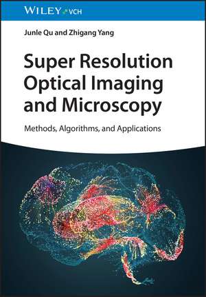
Super Resolution Optical Imaging and Microscopy
Editat de Junle Qu, Zhigang Yangen Limba Engleză Hardback – 31 ian 2024
Domeniul imagisticii optice a cunoscut o evoluție fulminantă de la stabilirea limitei de difracție, transformându-se dintr-o metodă de observație statică într-un instrument de nanoscopie dinamică. Super Resolution Optical Imaging and Microscopy – Methods, Algorithms, and Applications, publicată de Wiley Vch, reflectă această tranziție prin integrarea celor mai noi progrese în tehnicile SRM (Super-Resolution Microscopy). Apreciem în mod deosebit modul în care J Qu reușește să pună în echilibru rigoarea fizică a ingineriei funcției de răspuns la punct (PSF) cu nevoile practice ale chimiștilor care dezvoltă sonde fluorescente. Lucrarea acoperă aceeași arie tematică precum Super-Resolution Microscopy for Material Science de Lorenzo Albertazzi, însă abordarea este mult mai orientată către biologia aplicată și dezvoltarea de algoritmi. În timp ce alte titluri se concentrează pe fizica instrumentului, acest volum prioritizează criteriile de selecție a fluoroforilor și evaluarea critică a obstacolelor tehnice întâlnite în experimentele reale. Structura cărții urmărește o progresie logică: începe cu bazele SMLM (Single Molecule Localization Microscopy) și microscopia prin iluminare structurată, avansează către utilizarea rețelelor neuronale (Deep Learning) pentru îmbunătățirea imaginilor și culminează cu strategii inovatoare pentru evitarea marcajelor fluorescente (Label-Free SRM). Subliniem relevanța capitolului dedicat materialelor fluorescente, care servește drept ghid pentru cercetătorii ce urmăresc să depășească barierele actuale ale fotostabilității. Prin includerea protocoalelor de pregătire a probelor și a metodelor de procesare a imaginii, volumul devine o resursă indispensabilă pentru laboratoarele care adoptă tehnologii de nanoscopie.
Preț: 820.78 lei
Preț vechi: 1065.94 lei
-23%
Carte disponibilă
Livrare economică 10-16 noiembrie
Livrare express 31 octombrie-06 noiembrie pentru 80.61 lei
Specificații
ISBN-10: 3527349863
Pagini: 256
Ilustrații: 100 schwarz-weiße und 50 farbige Abbildungen
Dimensiuni: 172 x 246 x 19 mm
Greutate: 0.65 kg
Ediția:1
Editura: Wiley-VCH GmbH
Locul publicării:Weinheim, Germany
V-ar putea interesa
-
 Fundamentals of Micromechanics of SolidsJianmin Qu-9%Preț: 943.69 lei1037.02 lei
 -
 Artificial Intelligence and Spectroscopic Techniques for Gemology ApplicationsAshutosh Kumar Shukla-18%Preț: 1096.20 lei1336.83 lei
 -
 NukedLinda C MoricePreț: 157.74 lei
 -
 Microplastics in Urban Water ManagementBing-Jie Ni-9%Preț: 1070.07 lei1175.90 lei
 -
 -14%Preț: 1350.37 lei1570.19 lei
 -
 -9%Preț: 1091.60 lei1199.55 lei
 -
 Sodium-Ion BatteriesMaria-Magdalena Titirici-14%Preț: 1702.06 lei1979.14 lei
 -
 -14%Preț: 870.25 lei1011.92 lei
 -
 -14%Preț: 825.12 lei959.44 lei
 -
 -5%Preț: 1118.31 lei1177.18 lei
 -
 -14%Preț: 882.99 lei1026.74 lei
 -
 Rechargeable Ion BatteriesKaterina E. Aifantis-23%Preț: 922.94 lei1198.63 lei
 -
 ¿-cyclodextrine polymère dans les eaux usées textilesGurumeet WadhawaPreț: 283.12 lei
 -
 Polimero di ¿-ciclodestrina in acque reflue tessiliGurumeet WadhawaPreț: 282.87 lei
De ce să citești această carte
Recomandăm această lucrare cercetătorilor din chimie și biologie care utilizează microscopia avansată. Cititorul câștigă o înțelegere profundă a modului în care algoritmii de Deep Learning și designul sondelor fluorescente pot optimiza rezoluția la scară nanometrică. Este un instrument practic esențial pentru a naviga prin complexitatea pregătirii probelor și procesării datelor în studiile de super-rezoluție moderne.
Cuprins
Preface xi
1 Super-Resolution Microscopy (SRM): Brief Introduction 1
Zhigang Yang, Soham Samanta, and Yingchao Liu
1.1 Optical Microscopy 1
1.1.1 History and Background 1
1.2 Specialized Optical Microscopes 3
1.2.1 Inverted Microscopes 4
1.2.2 Confocal Microscopes 4
1.3 Optical Diffraction Limit 5
1.4 Super-Resolution Microscopy: Overcoming the Diffraction Limit 6
1.5 Near-Field Scanning Optical Microscopy 7
1.6 Far-Field Super-Resolution Microscopy 8
1.7 Fluorescent Probes for Super-Resolution Microscopy 9
1.8 Image Analysis Algorithms 10
1.9 Applications 11
1.10 Outline of the Content of Succeeding Chapters 11
Acknowledgment 11
References 12
2 Point Spread Function Engineering SRM 15
Wei Yan, Luwei Wang, Yinru Zhu, Jialin Wang, and Ruijie Xiang
2.1 Stimulated Emission Depletion Microscopy (STED) 15
2.1.1 Principles of STED 15
2.1.2 Three-Dimensional STED 16
2.1.3 Multi-Color and Multi-Photon STED 18
2.1.4 Strategies to Reduce STED Power 20
2.1.4.1 Time-Gated STED Technology 21
2.1.4.2 Offline Gated STED Technology 22
2.1.4.3 Phasor-Plot Analysis of STED-FLIM 23
2.1.4.4 STED Super-Resolution Imaging with Quantum Dots 24
2.1.4.5 Temporal and Spatial Modulation STED 26
2.1.4.6 STED Super-Resolution Imaging Based on Adaptive Optics 27
2.1.5 Live Cell Imaging 29
2.2 Ground State Depletion (GSD) Microscopy 32
2.2.1 Principles of GSD 32
2.2.2 Advantages and Disadvantages of GSD 33
2.2.3 Applications of GSD 34
2.3 Reversible Saturable Optical Fluorescence Transition Microscopy 34
2.3.1 Improvement in the RESOLFT System 36
2.3.1.1 Parallelized RESOLFT Microscopy 36
2.3.1.2 Two-Photon RESOLFT 37
2.3.1.3 Dual-Channel RESOLFT Imaging 37
2.3.1.4 Three-Dimensional Imaging 37
2.3.2 Fluorescent Probe for RESOLFT Microscopy 38
2.3.2.1 Early-Stage: Fluorescent Protein 38
2.3.2.2 Improvement Based on Fluorescence Dynamics 39
2.3.2.3 Improvement in Other Properties 39
2.3.2.4 Organic Fluorophores 41
2.3.3 Advances in RESOLFT Application 42
2.3.3.1 Application in Life Science 42
2.3.3.2 Application in Writing and Manufacturing at the Nanoscale 43
2.4 Conclusion 44
Acknowledgment 44
References 45
3 Single-Molecule Localization Microscopy (SMLM) 51
Danying Lin, Yingying Jing, Pengfa Chen, Zekai Wu, Zhenquan Gong, Jiao Zhang, Arup Tarai, and Xuehua Wang
3.1 Main Idea of SMLM 51
3.2 Stochastic Optical Reconstruction Microscopy (STORM) 53
3.2.1 Implementation of STORM 53
3.2.1.1 Typical Optical Setup 53
3.2.1.2 Two Key Steps 54
3.2.1.3 Derivative Forms 56
3.2.2 Key Consideration in STORM 57
3.2.3 Multi-Color STORM 59
3.2.4 Three-Dimensional STORM 61
3.2.4.1 PSF Engineering 63
3.2.4.2 Multi-Focal Plane Imaging 67
3.2.4.3 Other Methods 68
3.2.5 Live Cell STORM Imaging 69
3.3 Photo-Activated Localization Microscopy (PALM) 72
3.3.1 Basic Principle of PALM and Differences with STORM 72
3.3.2 Single-Particle Tracking PALM (sptPALM) 73
3.4 Point Accumulation for Imaging in Nanoscale Topography (paint) 75
3.4.1 Basic Principle, Advantages, and Disadvantages of PAINT 75
3.4.2 Modifications of PAINT 76
3.4.2.1 uPAINT 76
3.4.2.2 DNA-PAINT and Exchange-PAINT 76
3.5 Single-Molecule Localization Algorithms 78
3.5.1 Algebraic Algorithms 78
3.5.2 Single-Emitter Fitting Algorithms 79
3.5.3 Multi-Emitter Fitting Algorithms 80
3.5.4 CS Algorithms 82
3.5.5 Other Methods 83
3.6 Minflux 84
3.7 Conclusion 84
Acknowledgment 85
References 85
4 Fluorescence Fluctuation-Based Super-Resolution Imaging 93
Xuehua Wang and Bin Yu
4.1 Stochastic Optical Fluctuation Imaging (SOFI) 94
4.1.1 XC-SOFI 95
4.1.2 bSOFI 96
4.1.3 fSOFI 96
4.1.4 Speckle SOFI 97
4.2 Other Techniques 99
4.2.1 VISion 99
4.2.2 Bayesian Analysis of Blinking and Bleaching (3B) 99
4.2.3 Super-resolution Radial Fluctuations (SRRF) 100
4.2.4 Entropy-Based Super-Resolution Imaging (ESI) 101
4.2.5 Multiple Signal Classification Algorithm for Super-resolution Fluorescence Microscopy (MUSICAL) 102
4.3 Applications of Fluorescence Fluctuation-Based SRM Methods 102
4.4 Conclusion 103
Acknowledgment 104
References 104
5 Structured Illumination Microscopy 107
Bin Yu, Siwei Li, Faiz Wali, and Rong Xu
5.1 Introduction 107
5.2 Wide-field SIM 107
5.2.1 Basics of SIM 108
5.2.2 SR-SIM 110
5.2.2.1 Conventional Grating-Based SIM 111
5.2.2.2 Blind SIM 113
5.2.2.3 Grazing Incidence SIM (GI-SIM) 116
5.2.2.4 Hessian-SIM 117
5.2.3 Summary 118
5.3 Point-Scanning SIM 118
5.3.1 Principle of PS-SIM 119
5.3.2 PS-SIM Based on the Digital Method 121
5.3.3 PS-SIM Based on the Optical Method 123
5.3.4 Special PS-SIM 126
5.3.5 Summary 127
5.4 Conclusions and Future Prospects 128
Acknowledgement 129
References 129
6 Deep Learning-Based SR Microscopy 135
Jia Li and Jianhui Liao
6.1 Introduction 135
6.2 Fundamentals of Deep Networks 135
6.2.1 Neural Networks 136
6.2.2 Activation Function and Layers 137
6.2.2.1 Sigmoid 138
6.2.2.2 Softmax 139
6.2.2.3 Rectified Linear Unit (ReLU) 139
6.2.2.4 Leaky ReLU 140
6.2.3 Training and Data 141
6.2.3.1 Gradient Descent 141
6.2.3.2 Backpropagation 142
6.2.3.3 Data 143
6.2.4 Loss Functions 144
6.3 Deep Learning for SR Image Reconstruction 144
6.3.1 2D Reconstruction Methods 145
6.3.1.1 Convolutional Neural Networks (CNNs) 145
6.3.1.2 Convolutional Layer 146
6.3.1.3 Pooling Layer 147
6.3.1.4 Properties 147
6.3.1.5 SR Image Reconstruction with CNN 148
6.3.1.6 Generative Adversarial Networks (GANs) 149
6.3.1.7 Game Theory 150
6.3.1.8 Architecture 150
6.3.1.9 Training 150
6.3.1.10 SR Image Reconstruction with GAN 151
6.3.2 3D Reconstruction Methods 153
6.4 Challenges of Deep Learning-Based Methods 153
6.4.1 Data Limitations 154
6.4.2 Training Obstacles 154
6.4.3 Result Reliability 155
6.5 Conclusion 156
References 158
7 Fluorescent Materials for Super-Resolution Imaging 163
Zhigang Yang and Soham Samanta
7.1 Fluorescent Probes for Super-Resolution Imaging 163
7.2 Fluorescent Proteins 164
7.2.1 FPs for STED and RESOLFT Nanoscopy 164
7.2.2 FPs for SMLM-Based SRM 169
7.2.3 FPs for SIM and Other New SRM Techniques 176
7.3 Small-Molecule Fluorescent Probes 176
7.3.1 Organic Fluorescent Probes for STED 176
7.3.1.1 Rhodamine-Based Fluorescent Probes for STED Imaging 177
7.3.1.2 Diverse Fluorescent Probes for STED Imaging 179
7.3.1.3 Phosphole-Based Fluorescent Probes for STED Imaging 183
7.3.2 Organic Fluorescent Probes for SMLM 185
7.3.2.1 Xanthene/Rhodamine Dyes 185
7.3.2.2 Cyanine Dyes 191
7.3.2.3 BODIPY and Oxazine/Spiropyran Dyes 194
7.3.2.4 Other Dyes (2-dithienylethenes and Cicyanodihydrofurans) 198
7.3.3 Organic Fluorescent Probes for SIM 199
7.4 Fluorescent Metal Complexes for SRM 202
7.4.1 Fluorescent Metal Complexes for STED 202
7.4.2 Fluorescent Metal Complexes for SMLM 203
7.4.3 Fluorescent Metal Complexes for SIM 204
7.5 Fluorescent Nanomaterials (Nanoparticles/Quantum Dots/Carbon Nanotubes/Carbon Dots (CDs)/Polymers Dots) for SRM 204
7.5.1 Fluorescent Nanomaterials for STED 205
7.5.2 Organic Nanoparticles 205
7.5.3 Inorganic Nanoparticles 211
7.5.4 Fluorescent Nanomaterials for SMLM 213
7.5.5 Fluorescent Nanomaterials for SIM 216
Acknowledgment 218
References 219
8 Conclusion and Future Perspectives 229
Zhigang Yang, Soham Samanta, and Junle Qu
Index 235
Descriere scurtă
Descriere
Notă biografică
Dr. Junle Qu is a Professor in the College of Physics and Optoelectronic Engineering at Shenzhen University and the director of Center for Biomedical Optics and Photonics of Shenzhen University. His current research interests include biomedical optical imaging and imaging guided optical therapy. He has published more than 500 papers in peer reviewed journals.
Dr. Zhigang Yang is currently an associate Professor in the College of Physics and Optoelectronic Engineering, Shenzhen University and a member of Center for Biomedical Optics and Photonics of Shenzhen University. His research interests mainly focus on development of optical probes and biological imaging, including super-resolution fluorescent imaging, fluorescence lifetime imaging, and Visible-Near Infrared I/II imaging.