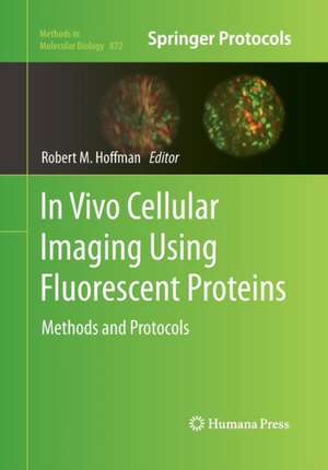
In Vivo Cellular Imaging Using Fluorescent Proteins
Editat de Robert Hoffmanen Limba Engleză Paperback – 23 aug 2016
Notăm cu interes faptul că actualizarea ghidurilor de cercetare biomedicală pune un accent tot mai mare pe observația neinvazivă, transformând proteinele fluorescente dintr-o curiozitate biologică într-un standard de aur pentru imagistica in vivo. Reținem că volumul In Vivo Cellular Imaging Using Fluorescent Proteins, editat de Robert Hoffman, reprezintă un punct de cotitură în înțelegerea proceselor celulare dinamice, oferind instrumentele necesare pentru a face vizibil ceea ce, până recent, era imperceptibil în interiorul unui organism viu. Structura cărții urmărește o progresie logică, de la bazele ingineriei genetice a proteinelor far-red și infraroșii, până la aplicații clinice complexe, precum monitorizarea angiogenezei tumorale și a răspunsului vascular la terapiile anti-cancer. Merită menționat că, spre deosebire de lucrările anterioare ale editorului, cum este Meeting Excellence care se concentra pe management organizațional, acest volum revine la rigoarea clinică și științifică prezentă în Goldfrank's Clinical Manual of Toxicologic Emergencies, Second Edition, aplicată de data aceasta în sfera oncologiei experimentale. Comparativ cu In Vivo Fluorescence Imaging de Mingfeng Bai, care oferă o perspectivă multidisciplinară asupra instrumentației, lucrarea de față este o alternativă esențială pentru cercetători și rezidenți datorită formatului său de tip „manual de laborator”. Avantajul major constă în includerea secțiunilor de „troubleshooting”, care permit reproducerea protocoalelor pentru modelele de metastaze peritoneale sau pleurale fără erorile metodologice comune. Este o resursă care documentează revoluția începută de proteinele fluorescente, oferind soluții tehnice pentru vizualizarea tridimensională a tumorilor în timp real.
Preț: 860.09 lei
Preț vechi: 905.36 lei
-5%
Carte tipărită la comandă
Livrare economică 03-17 iunie
Specificații
ISBN-10: 1493961497
Pagini: 284
Ilustrații: XIII, 269 p. 47 illus., 41 illus. in color.
Dimensiuni: 178 x 254 x 16 mm
Greutate: 0.54 kg
Ediția:Softcover reprint of the original 1st ed. 2012
Editura: Humana
Locul publicării:Totowa, NJ, United States
V-ar putea interesa
-
 Oxford Handbook of OncologyMiranda Payne-14%Preț: 282.45 lei328.19 lei
 -
 Oncologic Imaging: A Multidisciplinary ApproachPaul M. Silverman-29%Preț: 1174.39 lei1648.15 lei
 -
 Clinical Immuno-OncologyJohn E. Niederhuber-30%Preț: 717.47 lei1021.66 lei
 -
 Fluorescence Imaging and Biological QuantificationRaquel Seruca-5%Preț: 826.67 lei870.18 lei
 -
 Optical Imaging of CancerEben Rosenthal-5%Preț: 1063.94 lei1119.93 lei
 -
 Cellular and Molecular ImmunologyAbul K. Abbas-30%Preț: 475.59 lei677.09 leiRecomandat
 -
 -5%Preț: 1165.29 lei1226.62 lei
 -
 Pancreatic CancerHoward Reber-5%Preț: 1381.65 lei1454.37 lei
 -
 HypoxiaDaniele M. Gilkes-5%Preț: 1235.75 lei1300.79 lei
 -
 Breast CancerJian Cao-5%Preț: 700.70 lei737.57 lei
 -
 -15%Preț: 633.43 lei745.21 lei
 -
 -22%Preț: 557.41 lei716.03 lei
 -
 Cell Migration in Development, Health and DiseaseAnke Brüning-Richardson-15%Preț: 487.18 lei573.15 lei
 -
 Fluorescence Imaging for SurgeonsFernando D. Dip-5%Preț: 713.59 lei751.14 lei
 -
 Sensors and Probes for BioimagingY–T Chang-14%Preț: 788.80 lei917.20 lei
 -
 -5%Preț: 1889.61 lei1989.06 lei
 -
 -5%Preț: 1012.38 lei1065.66 lei
 -
 -5%Preț: 1082.71 lei1139.69 lei
 -
 Fluorescence-Guided SurgeryTakeaki Ishizawa-5%Preț: 1079.87 lei1136.71 lei
 -
 -24%Preț: 1308.23 lei1721.35 lei
De ce să citești această carte
Recomandăm această lucrare profesioniștilor din oncologie și biologie moleculară care doresc să implementeze tehnici de imagistică intravitală. Cititorul câștigă acces la protocoale verificate pentru monitorizarea metastazelor în modele animale, beneficiind de instrucțiuni clare pentru utilizarea lentivirusurilor și a adenovirulurilor în marcarea celulară. Este un instrument practic indispensabil pentru optimizarea studiilor preclinice de eficacitate terapeutică.
Despre autor
Robert Hoffman, editorul acestui volum, este o figură centrală în dezvoltarea metodologiilor de imagistică moleculară, ocupând poziția de director executiv în dezvoltare organizațională la Novartis Oncology. Experiența sa vastă în cercetarea cancerului și în managementul sistemelor bazate pe echipe de înaltă performanță se reflectă în rigoarea și claritatea acestui manual. Deși portofoliul său include titluri diverse, de la managementul întâlnirilor la manuale de urgențe toxicologice, contribuția sa în seria Methods in Molecular Biology subliniază angajamentul său față de avansul tehnic în vizualizarea proceselor biologice in vivo.
Descriere scurtă
Authoritative and easily accessible, In Vivo Cellular Imaging Using Fluorescent Proteins: Methods and Protocols is the first volume in the new field of in vivo cell biology and it serves both professionals and novices with its well-honed methodologies.
Cuprins
Recenzii
“Robert M. Hoffman introduces In vivo Cellular Imaging Using Fluorescent proteins, the eighteen chapters book dedicated to the description of how fluorescence proteins have changed the way to analyze cellular processes in vivo. … This book is very well structured with several colored figures … . this is a useful book not only for those who study the biology of different cancer cells, but also for those who are interested in following the in vivo dynamic of cellular processes.” (Manuela Monti, European Journal of Histochemistry, Vol. 56, 2012)