Essentials of Clinical Anatomy of the Equine Locomotor System
Autor Jean-Marie Denoixen Limba Engleză Hardback – 21 dec 2018
Preț: 526.12 lei
Preț vechi: 553.81 lei
-5% Nou
93.10€ • 108.57$ • 81.74£
Carte disponibilă
Livrare economică 25 decembrie 25 - 08 ianuarie 26
Livrare express 11-17 decembrie pentru 49.69 lei
Specificații
ISBN-10: 1498754414
Pagini: 308
Ilustrații: 231 Illustrations, color; 52 Illustrations, black and white
Dimensiuni: 174 x 256 x 25 mm
Greutate: 0.87 kg
Ediția:1
Editura: Taylor & Francis
Colecția CRC Press
Locul publicării:Boca Raton, United States
V-ar putea interesa
-
 -9%Preț: 1807.80 lei1986.60 lei
 -
 -5%Preț: 280.46 lei295.23 lei
 -
 Biomechanics and Physical Training of the HorseJean-Marie Denoix-5%Preț: 400.83 lei421.93 lei
 -
 Management of heat stress in broilers by organic feed supplementsMuhammad Umar Sohail-5%Preț: 356.83 lei375.61 lei
 -
 Vosproizvoditel'naya sposobnost' vysokoproduktivnykh korovLobodin Konstantin-5%Preț: 484.64 lei510.14 lei
 -
 Detección de infecciones por Trypanosoma vivaxAna Maria Bolivar-5%Preț: 173.59 lei182.73 lei
 -
 -5%Preț: 294.75 lei310.27 lei
 -
 -5%Preț: 355.98 lei374.71 lei
 -
 -5%Preț: 297.40 lei313.05 lei
 -
 Mycobacteria species in milk and some milk productWalaa Mahmoud Elsherif-5%Preț: 409.72 lei431.29 lei
 -
 -5%Preț: 242.16 lei254.90 lei
Public țintă
Adult education, Postgraduate, Professional, and Professional Practice & DevelopmentCuprins
Descriere
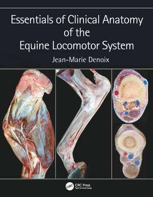
Essentials of Clinical Anatomy of the Equine Locomotor System presents a unique photographic record of dissections showing the topographical anatomy of the locomotor system of the horse. Readers of this book will be able to see the position and relationships of the bones, joints, muscles, nerves and blood vessels that make up each region of the forelimb, vertebral column and hindlimb.
Key features:
- Important features of regional and topographical anatomy are presented using full-color photos of detailed dissections
- Anatomy is presented in a clinical context
- Preparations of cross-sectional anatomy facilitate interpretation of diagnostic imaging, such as ultrasonography, MRI images and CT scans
- All dissections are of fresh material, rather than preserved specimens, to demonstrate the appearance of tissues in the living animal, or at post mortem autopsy
This new atlas is essential for anybody involved in detailed anatomical study, complex lameness evaluation or advanced imaging techniques in horses. It will be a useful guide for veterinary students, and a reference for equine vets in practice.