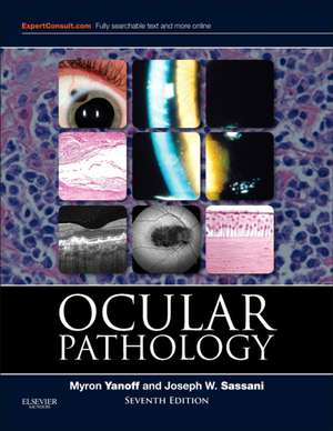
Ocular Pathology
Autor Myron Yanoff, Joseph W. Sassanien Limba Engleză Hardback – 19 mai 2014
Ne-a atras atenția, în primul rând, rigurozitatea componentelor vizuale din această a noua ediție a Ocular Pathology. Tabelele de diagnostic diferențial și figurile clinice nu servesc doar ca material ilustrativ, ci constituie piloni de referință pentru diagnosticul morfologic. Notăm cu interes modul în care autorii, Myron Yanoff și Joseph W. Sassani, reușesc să mențină un echilibru între concizia necesară practicii clinice și profunzimea academică cerută de un tratat de referință.
Merită menționat că volumul este organizat într-o succesiune logică, pornind de la principiile fundamentale ale patologiei (inflamație, imunobiologie, reacții tisulare) și avansând spre entități specifice. Capitolele dedicate anomaliilor congenitale, uveitei și endoftalmitei sunt completate de o secțiune extinsă despre traumele chirurgicale și non-chirurgicale. Această structură permite o navigare rapidă către complicațiile specifice intervențiilor pe cornee sau retină. Pe linia clinică a lucrării Lee's Ophthalmic Histopathology, dar cu accent pe implementarea datelor genetice și a tehnicilor de imagistică de ultimă oră, volumul de față ancorează patologia oculară în medicina contemporană.
Poziționarea autorului Myron Yanoff în literatura de specialitate este una centrală, acesta fiind și coordonatorul seriei Advances in Ophthalmology and Optometry. Dacă lucrările sale anterioare se concentrau pe actualizări anuale ale practicii, Ocular Pathology rămâne fundamentul teoretic care explică mecanismele bolii. Această ediție actualizează nosologia oculară și metodele de clasificare, fiind o resursă indispensabilă pentru înțelegerea evoluției conceptelor curente în morfologia oculară.
Preț: 1400.85 lei
Preț vechi: 1998.81 lei
-30%
Carte disponibilă
Livrare economică 21 august-02 septembrie
Specificații
ISBN-10: 1455728748
Pagini: 714
Ilustrații: Approx. 1917 illustrations (1729 in full color)
Dimensiuni: 222 x 282 x 40 mm
Greutate: 2.31 kg
Ediția:7th Edition.
Editura: Elsevier
V-ar putea interesa
-
 The Retinal AtlasK. Bailey Freund-28%Preț: 1738.34 lei2412.96 lei
 -
 -39%Preț: 377.82 lei618.19 lei
 -
 -5%Preț: 1040.84 lei1095.61 lei
 -
 OphthalmologyMyron Yanoff-27%Preț: 1817.39 lei2498.40 lei
 -
 Atlas of Retinal OCT: Optical Coherence TomographyJay S. Duker-29%Preț: 1000.41 lei1410.68 lei
 -
 Video Atlas of Ophthalmic UltrasoundBernadete Ayres-29%Preț: 1006.45 lei1416.46 lei
 -
 Kanski's Clinical Ophthalmology: A Systematic ApproachJohn F. Salmon-28%Preț: 1219.48 lei1683.23 lei
 -
 Clinical Cases in Ocular Oncology: Differential Diagnosis and ManagementAparna Ramasubramanian-30%Preț: 581.98 lei827.43 lei
 -
 -28%Preț: 791.24 lei1104.08 leiRecomandat
 -
 -23%Preț: 663.95 lei857.57 lei
 -
 -5%Preț: 550.14 lei579.10 lei
 -
 Kanski's Synopsis of Clinical OphthalmologyJohn F. Salmon-28%Preț: 497.77 lei693.12 lei
 -
 Clinical Atlas of Canine and Feline Ophthalmic DiseaseDouglas Esson-5%Preț: 1198.43 lei1261.50 lei
 -
 Uveitis: Fundamentals and Clinical Practice: Expert Consult - Online and PrintRobert B. Nussenblatt-29%Preț: 852.36 lei1202.93 lei
 -
 -25%Preț: 2715.37 lei3632.23 lei
 -
 -5%Preț: 703.43 lei740.45 lei
 -
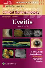 UveitisSunir J. Garg, MD-27%Preț: 579.31 lei792.63 lei
 -
 Clinical OphthalmologyMarieta Dumitrache-5%Preț: 1386.36 lei1459.32 lei
 -
 Pocket Guide to Ocular Oncology and PathologyHans Grossniklaus-5%Preț: 1087.82 lei1145.08 lei
 -
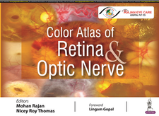 Color Atlas of Retina & Optic NerveMohan Rajan-5%Preț: 659.96 lei694.69 lei
De ce să citești această carte
Recomandăm această lucrare oftalmologilor și patologilor care caută o bază factuală solidă pentru diagnostic. Ediția a 9-a aduce noutăți esențiale în genetică și imagistică, elemente care transformă modul în care înțelegem patogeneza oculară. Este un instrument clinic precis, premiat de British Medical Association, care facilitează corelația dintre observația clinică și substratul histologic.
Descriere scurtă
"This seventh edition of Ocular Pathology by Myron Yanoff and Joseph Sassani is a superb update of what has become the single best ophthalmic pathology reference text for ophthalmologists, pathologists and researchers."Foreword by:J. Douglas Cameron, Ophthalmology and Visual Neurosciences, University of Minnesota School of Medicine, June 2015
Cuprins
Inflammation
Immunobiology
Cellular and Tissue Reactions
2. CONGENITAL ANOMALIES
Phakamatoses (Disseminated Hereditary Hamartomas)
Chromosomal Aberrations
Infectious Embryopathy
Drug Embryopathy
Other Congenital Anomalies
3. NONGRANULOMATOUS INFLAMMATION:
UVEITIS, ENDOPHTHALMITIS, PANOPHTHALMITIS, AND SEQUELAE
Classification
Suppurative Endophthalmitis and Panophthalmitis
Nonsuppurative, Chronic Nongranulomatous Uveitis and Endophthalmitis
Sequelae of Uveitis, Endophthalmitis, and Panophthalmitis
End Stage of Diffuse Ocular Diseases
4. GRANULOMATOUS INFLAMMATION
Introduction
Post-Traumatic
Nontraumatic Infectious
Nontraumatic Noninfectious
5. SURGICAL AND NONSURGICAL TRAUMA
Causes of Enucleation
Normal Wound Healing
Complications of Intraocular Surgery
Complications of Neural Retinal Detachment and Vitreous Surgery
Complications of Corneal Surgery
Complications of Nonsurgical Trauma
6. SKIN AND LACRIMAL DRAINAGE SYSTEM
SKIN
Normal Anatomy
Terminology
Congenital Abnormalities
Aging
Inflammation
Lid Manifestations of Systemic Dermatoses or Disease
Cysts, Pseudoneoplasms, and Neoplasms
LACRIMAL DRAINAGE SYSTEM
Normal Anatomy
Congenital Abnormalities
Inflammation - Dacryocystitis
Tumors
7. CONJUNCTIVA
Normal Anatomy
Congenital Anomalies
Vascular Disorders
Inflammation
Injuries
Conjunctival Manifestations of Systemic Disease
Degenerations
Cysts, Pseudoneoplasms, and Neoplasms
8. CORNEA AND SCLERA
CORNEA
Normal Anatomy
Congenital Defects
Inflammations - Nonulcerative
Inflammations - Ulcerative
Inflammations - Corneal Sequelae
Injuries
Degenerations
Dystrophies
Pigmentations
SCLERA
Congenital Anomalies
Inflammations
Injuries
Tumors
9. UVEA
Normal Anatomy
Congenital and Developmental Defects
Congenital and Developmental Defects of the Pigment Epithelium
Inflammations
Injuries Systemic Diseases
Atrophies and Degenerations
Dystrophies
Tumors Uveal Edema
10. LENS
Normal Anatomy
General Information
Congenital Anomalies
Capsule (Epithelial Basement Membrane)
Epithelium
Cortex and Nucleus (Lens Cells or "Fibers)
Secondary Cataracts
Complications of Cataracts
Ectopic Lens
11. NEURAL (SENSORY) RETINA
Normal Anatomy
Congenital Anomalies
Vascular Diseases
Inflammations
Injuries
Degenerations
Hereditary Primary Retinal Dystrophies
Hereditary Secondary Retinal Dystrophies
Systemic Diseases Involving the Retina
Tumors
Neural Retinal Detachment
12. VITREOUS
Normal Anatomy
Congenital Anomalies
Inflammation
Vitreous Adhesions
Vitreous Opacities
Vitreous Hemorrhage
13. OPTIC NERVE
Normal Anatomy
Congenital Defects and Anatomic Variations
Optic Disc Edema
Optic Neuritis
Optic Atrophy
14. ORBIT
Normal Anatomy
Exophthalmos
Developmental Abnormalities
Orbital Inflammation
Injuries
Vascular Disease
Ocular Muscle Involvement in Systemic Disease
Neoplasms and Other Tumors
15. DIABETES MELLITUS
Natural History
Retinal Vasculature in Normals and Diabetics
Conjunctiva and Cornea
Lens
Iris
Ciliary Body and Choroid
Neurosensory Retina
Vitreous
Optic Nerve
16. GLAUCOMA
Normal Anatomy
Introduction
Normal Outflow
Tissue Changes Caused by Elevated Intraocular Pressure
17. OCULAR MELANOTIC TUMORS
Normal Anatomy
Melanotic Tumors of Eyelids
Melanotic Tumors of Conjunctiva
Melanotic Tumors of Pigment Epithelium of Iris, Ciliary Body, and Retina
Melanotic Tumors of the Uvea
Melanotic Tumors of the Optic Disc
Melanotic Tumors of the Orbit
18. RETINOBLASTOMA AND PSEUDOGLIOMA
Retinoblastoma
Pseudoglioma