Cantitate/Preț
Produs

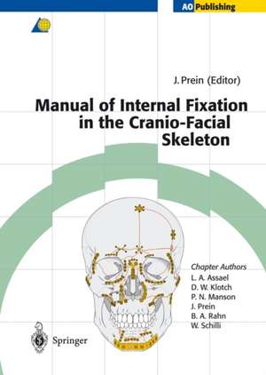
Manual of Internal Fixation in the Cranio-Facial Skeleton: Techniques Recommended by the AO/ASIF Maxillofacial Group

Editat de Joachim Prein Contribuţii de L.A. Assael, D.W. Klotch, P.N. Manson, J. Prein, B.A. Rahn, W. Schilli
en Limba Engleză Paperback – 23 aug 2014

Preț: 55742 lei

Preț vechi: 58676 lei
-5%

Puncte Express: 836

Preț estimativ în valută:
10680 11631$ 9120£

Carte tipărită la comandă

Livrare economică 26 aprilie-02 mai

Preluare comenzi: 021 569.72.76

Specificații

ISBN-13: 9783642637322
ISBN-10: 3642637329
Pagini: 244
Ilustrații: XIV, 227 p. 275 illus., 272 illus. in color.
Dimensiuni: 193 x 270 x 20 mm
Greutate: 0 kg
Ediția:Softcover reprint of the original 1st ed. 1998
Editura: Springer Berlin, Heidelberg
Colecția Springer
Locul publicării:Berlin, Heidelberg, Germany

Public țintă

Professional/practitioner

Cuprins

1 Scientific Background.- 1.1 Introduction.- 1.1.1 The AO/ASIF Foundation.- 1.1.2 Research.- 1.1.3 Development.- 1.1.4 Education.- 1.1.5 Documentation and Clinical Investigations.- 1.1.6 Fracture Classification.- 1.2 Bone as a Material.- 1.2.1 Structure.- 1.2.2 Chemical Composition.- 1.2.3 Mechanical Properties.- 1.2.4 Mechanical Glossary.- 1.3 Fractures in the Cranio-Maxillo-Facial Skeleton.- 1.3.1 Origin of Skull Bones.- 1.3.2 Load-Bearing Structures in the Cranio-Maxillo-Facial Skeleton.- 1.3.3 The Fracture.- 1.3.4 Biological Reaction and Healing of Bone.- 1.4 Indications for Operative Treatment of Fractures.- 1.5 Operative Reduction and Internal Fixation.- 1.5.1 Reestablishing Stability.- 1.5.2 Implant Materials.- 1.5.2.1 Stainless Steel.- 1.5.2.2 Titanium.- 1.5.2.3 Biodegradable Polymeric Materials.- 1.5.3 Implant Removal.- 1.5.4 Principles of Stabilization.- 1.5.4.1 Splinting.- 1.5.4.2 Compression.- 1.6 Design and Function of Implants and Instruments.- 1.6.1 Screws.- 1.6.1.1 Function of Screws.- 1.6.1.2 Types of Screws.- 1.6.1.3 List of Screws.- 1.6.1.4 Technique of Screw Insertion.- 1.6.1.5 Instruments for Screw Insertion.- 1.6.2 Plates.- 1.6.2.1 Craniofacial Plates.- 1.6.2.2 Mandible and Reconstruction Plates.- 1.6.3 Instruments.- 1.6.4 Power Tools.- 1.7 Set Configurations.- 1.7.1 European Set Configuration.- 1.7.2 North American Set Configuration.- 1.8 External Fixation Devices.- References and Suggested Reading.- 2 Anatomic Approaches.- 3 Mandibular Fractures.- 3.1 Introduction.- 3.2 Treatment Planning.- 3.3 Cost Effectiveness.- 3.4 Adequate Stability.- 3.5 Mistakes in Application and Technique.- 3.6 Failures.- 3.7 Indications for Osteosynthesis.- 3.8 Indications for Perioperative Antibiotic Cover.- 3.9 General Remarks.- 3.10 Localization and Types of Fracture.- 3.11 Fractures of the Symphysis and the Parasymphyseal Area.- 3.11.1 Tr ansverse Fracture Line Without Dislocation.- 3.11.2 Transverse Fracture Line With Dislocation.- 3.11.3 Fracture Line With Basal Triangle.- 3.11.4 Comminuted Fractures.- 3.12 Fractures of the Horizontal Ramus.- 3.12.1 Transverse Fracture Line Without Dislocation.- 3.12.2 Transverse Fracture Line With Dislocation.- 3.12.3 Oblique Fracture Line With/Without Dislocation.- 3.12.4 Fracture Line With Basal Triangle.- 3.12.5 Comminuted Fractures.- 3.13 Fractures of the Mandibular Angle.- 3.13.1 Transverse Fracture Line Without Dislocation.- 3.13.2 Transverse Fracture Line With Dislocation.- 3.13.3 Angular Fractures With Basal Triangle.- 3.13.4 Comminuted Angular Fractures.- 3.13.5 Comminuted Fractures of the Ascending Mandibular Ramus.- 3.14 Condylar and Subcondylar Fractures.- 3.14.1 Transverse Fracture Line With Dislocation.- 3.15 Fractures of the Atrophic Mandible.- 3.16 Infected Fractures.- 3.17 Defect Fractures.- 3.18 Mandibular Fractures in Children.- References and Suggested Reading.- 4 Craniofacial Fractures.- 4.1 Organization of Treatment in Panfacial Fractures.- 4.1.1 Introduction.- 4.1.2 Surgical Sequencing of Le Fort Fracture Treatment.- 4.1.3 Occlusion.- 4.1.4 Upper Face: The Cranial Unit.- 4.1.5 Upper Midfacial Unit.- 4.1.6 Lower Face.- 4.1.7 Linking the Upper and Lower Face.- 4.1.8 Edentulous Fractures.- 4.1.9 Soft Tissue.- 4.1.10 Soft-Tissue Injury.- 4.1.11 The “Double Insult” to Soft Tissue.- References and Suggested Reading.- 4.2 Le Fort I-III Fractures.- 4.2.1 Lower Midface (Le Fort I).- 4.2.1.1 Anatomy.- 4.2.1.2 Classification.- 4.2.1.3 Diagnosis.- 4.2.1.4 Le Fort I Fractures.- Treatment.- Airway Management.- Exposure.- Reduction.- Occlusion.- Fixation.- Bone Grafting.- Palatal Fractures.- Alveolar Ridge.- 4.2.2 Upper Midface (Le Fort II and III).- 4.2.2.1 Anatomy.- 4.2.2.2 Diagnosis.- 4.2.2.3 Treatment.- References and Suggested Reading.- 4.3 Naso-Orbital-Ethmoid Fractures.- 4.3.1 Definition.- 4.3.2 Anatomy.- 4.3.3 Fracture Patterns.- 4.3.4 Plates Used for Internal Fixation of NOE Fractures.- 4.3.5 Exposure.- 4.3.6 Diagnosis.- 4.3.7 Operative Treatment.- 4.3.7.1 Management of Central Fragment.- 4.3.7.2 Transnasal Canthopexy.- 4.3.7.3 Nasal Reconstruction.- 4.3.8 NOE Fracture-Related Problems.- 4.3.8.1 Lacrimal Duct Injuries.- 4.3.8.2 Frontal Sinus.- 4.3.8.3 Skull Base Injuries.- 4.3.8.4 Orbital Reconstruction.- References and Suggested Reading.- 4.4 Zygomatic Complex Fractures.- 4.4.1 Definition.- 4.4.2 Treatment.- 4.4.3 Exposure.- 4.4.4 Reduction.- 4.4.5 Stabilization.- 4.4.6 Internal Orbit.- 4.4.7 Soft-Tissue Closure.- References and Suggested Reading.- 4.5 Orbital Fractures.- 4.5.1 Definition.- 4.5.2 Diagnosis.- 4.5.3 Treatment.- 4.5.4 Reconstruction of Ethmoid Defects.- 4.5.5 Zygomatic (Lateral and Inferior Wall) Injuries.- 4.5.6 Naso-Orbital-Ethmoid Fractures.- 4.5.7 Superior Orbital Rim and Roof Fractures.- References and Suggested Reading.- 4.6 Cranial Vault.- 4.6.1 Frontal Sinus and Frontal Bone.- 4.6.2 Special Conditions Influencing Open Reduction and Internal Fixation.- 4.6.3 Sinus Function and Operative Treatment.- 4.6.4 Types of Fixation.- 4.6.5 Esthetics.- 4.6.6 Osteotomy.- 4.6.7 Exposure.- References and Suggested Reading.- 5 Reconstructive Tumor Surgery in the Mandible.- 5.1 Diagnosis.- 5.2 Patient Selection.- 5.3 Description of Procedures.- 5.3.1 Mandibular Osteotomy.- 5.3.2 Stabilization of Curetted Mandibular Defects.- 5.3.3 Segmental Resection for Benign Tumors.- 5.3.4 Plate Application for Tumors with Extension Through the Anterior Buccal Cortex.- 5.3.4.1 Plate Application for Anterior Tumor Extension Using the Mandible Fix Bridge Device.- 5.3.4.2 Fixation With Miniplates.- 5.3.4.3 Fixation With a Reconstruction Plate.- 5.3.4.4 Three-Dimensional Computer Modeling.- 5.3.5 Application of Bone Grafts Following Bridging Plate Stabilization.- 5.3.6 Reconstruction of Tumor Defects With Vascularized Bone Grafts and Their Fixation With Plates.- 5.3.7 Repair of the Anterior Defect Using a Microvascular Free Bone Flap and Fixation With Universal Fracture Plates (Microvascular Plates), UniLOCK, or Mini Plates.- 5.3.8 Repair of Tumor Defects With Anterior Soft-Tissue Extension with Microvascular Free Bone Flap and Bridging Plate.- 5.3.9 Reconstruction of the Condyle: General Remarks.- 5.3.10 Joint Repair With Costochondral Grafts.- 5.3.11 Alloplastic Replacement of the Condylar Process.- 5.3.12 Condylar Prosthesis for Lateromandibular Defects Including the Joint.- 5.3.13 Management of Mandibular Resection Including the Condyle Using Microvascular Bone Flaps and Various Plates for Fixation.- 5.3.14 Repair of the Lateral Mandible Including the Condyle: Osseous Free Flap and Bridging Plate.- 5.3.15 Repair of the Lateral Mandible and Condyle: Microvascular Free Flap and Reconstruction Plate With Condylar Prosthesis (Schusterman).- 5.3.16 Condylar Reconstruction: Condylar Transplant and Vascularized Bone Flap Reconstruction.- 5.4 Complications.- 5.5 Technical Errors.- 5.5.1 Plate Failure.- 5.5.2 Screw Failure.- 5.5.3 Soft-Tissue Failure.- 5.5.4 Joint Failure.- 5.5.5 Bone Failure.- References and Suggested Reading.- 6 Stable Internal Fixation of Osteotomies of the Facial Skeleton.- 6.1 Introduction.- 6.2 Treatment Planning for Internal Fixation of Osteotomies.- 6.3 Surgical Procedures.- 6.3.1 Mandibular Surgery.- 6.3.1.1 Sagittal Split Osteotomy.- 6.3.1.2 Other Ramus Osteotomies.- 6.3.1.3 Genioplasty.- 6.3.1.4 Mandibular Segmental Surgery.- 6.3.2 Midface Surgery: Le Fort I Osteotomy.- 6.4 Evaluation of Outcomes.- 6.5 Complications.- 6.6 Summary.- References and Suggested Reading.- 7 Craniofacial Deformities.- 7.1 Introduction.- 7.2 Incisions for Craniofacial Reconstruction and Patient Positioning.- 7.3 Craniosynostosis.- 7.4 Planning and Reconstruction.- 7.5 Surgical Technique: Anterior Cranial Expansion and Reconstruction.- 7.6 Posterior Cranial Expansion.- 7.7 Complete or Subtotal Calvarial Expansion.- 7.8 Hypertelorism.- 7.9 Monoblock Osteotomies.- 7.10 Orbital Dystopia.- 7.11 Craniofacial (Hemifacial) Microsomia.- 7.12 The Treacher Collins Malformation.- 7.13 Encephaloceles.- 7.14 Bone Lengthening by Continuous Distraction.- 7.14.1 Distraction for Mandibular Deformities.- 7.14.2 Midface Distraction.- References and Suggested Reading.

Caracteristici

Recent advances in fixation techniques of the bones in the cranium and the face * detailed guidelines as to operate on fractures, tumour defects, and osteotomies * surgical techniques as recommended by the AO/ASIF Maxillofacial Group* clear, detailed and instructive drawings *clinical situations shown on x-rays.