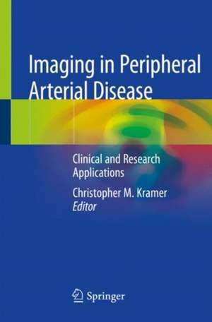
Imaging in Peripheral Arterial Disease
Editat de Christopher M. Krameren Limba Engleză Paperback – 7 oct 2020
Preț: 597.99 lei
Preț vechi: 629.46 lei
-5%
Puncte Express: 897
Carte tipărită la comandă
Livrare economică 17-23 noiembrie
Livrare prin curier în România Termenul estimat este afișat lângă disponibilitate.
Transport gratuit pentru acest produs Plată online sau ramburs, în funcție de opțiunile comenzii.
Retur gratuit în 14 zile Comandă securizată și suport în română.
Specificații
ISBN-13: 9783030245986
ISBN-10: 3030245985
Pagini: 236
Ilustrații: X, 223 p. 91 illus., 34 illus. in color.
Dimensiuni: 155 x 235 x 13 mm
Greutate: 0.41 kg
Ediția:1st edition 2020
Editura: Springer
Locul publicării:Cham, Switzerland
ISBN-10: 3030245985
Pagini: 236
Ilustrații: X, 223 p. 91 illus., 34 illus. in color.
Dimensiuni: 155 x 235 x 13 mm
Greutate: 0.41 kg
Ediția:1st edition 2020
Editura: Springer
Locul publicării:Cham, Switzerland
V-ar putea interesa
-
 -5%Preț: 1080.39 lei1137.25 lei
 -
 Investigating and Managing Common Cardiovascular ConditionsJuan Carlos Kaski-5%Preț: 364.38 lei383.56 lei
 -
 -5%Preț: 89.58 lei94.29 lei
 -
 -23%Preț: 641.35 lei836.74 lei
 -
 -23%Preț: 926.38 lei1209.59 lei
 -
 -23%Preț: 1210.93 lei1582.15 lei
 -
 Endoluminale VarizentherapieFlorian Johannes Netzer-5%Preț: 1338.75 lei1409.21 lei
 -
 Hypertension and Brain DamageAntonio Coca-5%Preț: 695.65 lei732.26 lei
 -
 Percutaneous Treatment of Left Side Cardiac ValvesCorrado Tamburino-5%Preț: 862.35 lei907.74 lei
 -
 Cancer and Cardiovascular Disease: A Concise Clinical AtlasSyed Wamique Yusuf-5%Preț: 1268.54 lei1335.31 lei
 -
 Essentials of HypertensionFlávio Danni Fuchs-5%Preț: 659.54 lei694.25 lei
 -
 Osteocardiology: Cardiac Bone FormationNalini M. Rajamannan-5%Preț: 352.64 lei371.20 lei
 -
 Anticoagulation TherapyJoe F. Lau-5%Preț: 1499.40 lei1578.31 lei
 -
 Cardiac Emergencies in ChildrenAshok P. Sarnaik-5%Preț: 1213.30 lei1277.15 lei
 -
 Right Heart PathologySilviu Ionel Dumitrescu-5%Preț: 2628.15 lei2766.48 lei
 -
 Thoracic Aortic DiseasesHervé Rousseau-5%Preț: 1023.73 lei1077.61 lei
 -
 -5%Preț: 278.91 lei293.58 lei
 -
 The Origin and Onset of Thrombus DiseaseLemin Wang-5%Preț: 350.93 lei369.40 lei
 -
 CLINIC ELECTROCARDIO (4TH ED)Chia B L-5%Preț: 195.14 lei205.41 lei
Cuprins
Part I. Clinical Applications.- Chapter 1. Imaging Needs of Clinicians Caring for PAD Patients.- Chapter 2. Role of the Ankle Brachial Index
Chapter 3. Duplex Ultrasound.- Chapter 4. Computed Tomography Angiography (CTA).- Chapter 5. Magnetic Resonance Angiography (MRA).- Chapter 6. Digital Subtraction Angiography.- Part II. Research Applications.- Chapter 7. Imaging Needs for Novel Therapeutics in PAD
Chapter 8. Contrast Ultrasound.- Chapter 9. MR Perfusion and Spectroscopy.- Chapter 10. MR Plaque Imaging.- Chapter 11. Radionuclide Imaging.
Notă biografică
Christopher M. Kramer, MD
Ruth C. Heede Professor of Cardiology
Professor of Radiology
Director,
University of Virginia Health System
Charlottesville, VA, 22908
USA
Ruth C. Heede Professor of Cardiology
Professor of Radiology
Director,
University of Virginia Health System
Charlottesville, VA, 22908
USA
Textul de pe ultima copertă
This book presents up-to-date information on clinical and research applications of imaging in peripheral arterial disease (PAD). It provides high-quality images useful not only in the diagnosis of PAD but also for use in clinical trials aimed at the development of novel therapies such as angiogenic agents and stem cells. The book begins with coverage of the applications of the four major imaging modalities in a clinical setting: ultrasound, computed tomography angiography (CTA), magnetic resonance angiography (MRA), and digital subtraction angiography (DSA). It also discusses the ankle brachial index (ABI) as a screening technique to establish the presence of PAD. Subsequent chapters focus on the advantages and limitations of various research applications of imaging in PAD including contrast ultrasound for measuring perfusion; MRI for assessing perfusion, energetics, plaque volume, and characteristics; and radionuclide imaging for perfusion and inflammation. Imaging in Peripheral Arterial Disease: Clinical and Research Applications is an essential resource for physicians, researchers, residents, and fellows in cardiology, radiology, imaging, nuclear medicine, diagnostic radiology, and vascular surgery.
Caracteristici
Focuses specifically on diagnosis. Covers clinical applications of ultrasound, CTA , MRA, and radionuclide imaging. Discusses research applications of contrast echo, magnetic resonance perfusion and spectroscopy, and radionuclide imaging. Highlights ankle brachial index (ABI) as a screening technique to establish the presence of PAD