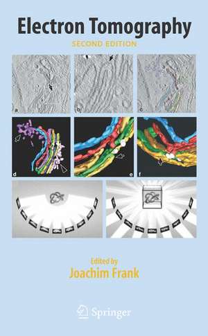
Electron Tomography
Editat de Joachim Franken Limba Engleză Hardback – 18 dec 2006
This extensively revised second edition updates key contributions on the mathematics of 3D reconstruction, and includes new topics such as automated tomography, frozen sectioning of cells, and the interpretation of density maps through methods of fitting, docking, denoising, and segmentation. Each chapter is a self-contained treatise by a world expert in the author's field of research, resulting in an indispensable resource and companion for laboratories that practice electron tomography or seek to implement electron tomography as a tool for visualization of cells and cell components.
Key Features
- Presents the mathematical background and working methods for three-dimensional reconstruction from projections
- Takes the reader from biological specimen preparation to three-dimensional images of the cell and its components
- Revised and updated extensively from the first edition published in 1992
- The definitive work in the field written by leading international experts
Preț: 1350.08 lei
Preț vechi: 1646.44 lei
-18%
Puncte Express: 2025
Carte tipărită la comandă
Livrare economică 09-23 septembrie
Livrare prin curier în România Termenul estimat este afișat lângă disponibilitate.
Transport gratuit pentru acest produs Plată online sau ramburs, în funcție de opțiunile comenzii.
Retur gratuit în 14 zile Comandă securizată și suport în română.
Specificații
ISBN-13: 9780387312347
ISBN-10: 038731234X
Pagini: 480
Ilustrații: XIV, 456 p. 123 illus.
Dimensiuni: 160 x 241 x 35 mm
Greutate: 0.97 kg
Ediția:2nd edition 2006
Editura: Springer
Locul publicării:New York, NY, United States
ISBN-10: 038731234X
Pagini: 480
Ilustrații: XIV, 456 p. 123 illus.
Dimensiuni: 160 x 241 x 35 mm
Greutate: 0.97 kg
Ediția:2nd edition 2006
Editura: Springer
Locul publicării:New York, NY, United States
V-ar putea interesa
-
 Nature's Robots: A History of ProteinsCharles Tanford-6%Preț: 201.49 lei213.31 lei
 -
 Protein Structure and FunctionGregory Petsko-22%Preț: 508.77 lei653.32 lei
 -
 Flexible VirusesVladimir Uversky-5%Preț: 1278.03 lei1345.30 lei
 -
 Introduction to Protein StructureCarl Ivar Branden-9%Preț: 697.95 lei766.98 lei
 -
 Computational and StatisticalIngvar Eidhammer-8%Preț: 684.48 lei744.00 lei
 -
 Biotechnology of Plasma ProteinsRoger L. Lundblad-18%Preț: 1478.51 lei1803.06 lei
 -
 -15%Preț: 648.56 lei763.01 lei
 -
 New Developments in Proteomics ResearchGoro Fukushima-30%Preț: 1156.05 lei1659.32 lei
 -
 New Research on Molecular ChaperonesJaime Wyatt-27%Preț: 985.61 lei1359.12 lei
 -
 Protein-Protein ComplexesMartin Zacharias-15%Preț: 496.66 lei584.30 lei
 -
 G Protein-Coupled Receptors - Modeling and SimulationMarta Filizola-18%Preț: 919.16 lei1120.92 lei
Public țintă
ResearchCuprins
Introduction: Principles of Electron Tomography.- Sample Shrinkage and Radiation Damage of Plastic Sections.- Electron Tomography of Frozen-hydrated Sections of Cells and Tissues.- The Electron Microscope as a Structure Projector.- Cryotomography: Low-dose Automated Tomography of Frozen-hydrated Specimens.- Fiducial Marker and Hybrid Alignment Methods for Single- and Double-axis Tomography.- Markerless Alignment in Electron Tomography.- Algorithms for Three-dimensional Reconstruction From the Imperfect Projection Data Provided by Electron Microscopy.- Weighted Back-projection Methods.- Reconstruction With Orthogonal Functions.- Resolution in Electron Tomography.- Denoising of Electron Tomograms.- Segmentation of Three-dimensional Electron Tomographic Images.- Segmentation of Cell Components Using Prior Knowledge.- Motif Search in Electron Tomography.- Localization and Classification of Repetitive Structures in Electron Tomograms of Paracrystalline Assemblies.
Textul de pe ultima copertă
Electron tomography has become a standard technique with applications in cell biology, structural biology, and materials science. This definitive work provides a comprehensive treatment of the mathematical background and working methods of three-dimensional reconstruction from tilt series, with special emphasis on the problems presented by limitations of data collection in the transmission electron microscope. In addition to chapters that are applicable to 3D reconstruction in all fields of science, such as radiological imaging in medicine and electron tomography in materials science, Electron Tomography also focuses on specimen preparation and imaging unique to biological electron microscopy.
This extensively revised second edition updates key contributions on the mathematics of 3D reconstruction, and includes new topics such as automated tomography, frozen sectioning of cells, and the interpretation of density maps through methods of fitting, docking, denoising, and segmentation. Each chapter is a self-contained treatise by a world expert in the author's field of research, resulting in an indispensable resource and companion for laboratories that practice electron tomography or seek to implement electron tomography as a tool for visualization of cells and cell components.
This extensively revised second edition updates key contributions on the mathematics of 3D reconstruction, and includes new topics such as automated tomography, frozen sectioning of cells, and the interpretation of density maps through methods of fitting, docking, denoising, and segmentation. Each chapter is a self-contained treatise by a world expert in the author's field of research, resulting in an indispensable resource and companion for laboratories that practice electron tomography or seek to implement electron tomography as a tool for visualization of cells and cell components.
Caracteristici
Presents the mathematical background and working methods for three-dimensional reconstruction from projections Takes the reader from biological specimen preparation to three-dimensional images of the cell and its components Revised and updated extensively from the first edition published in 1992 The definitive work in the field written by leading international experts