Colorectal Surgery
Autor Mark Killingbacken Limba Engleză Paperback – 18 dec 2008
Bazat pe o experiență clinică de peste trei decenii și pe observații directe din sala de operație, Colorectal Surgery reprezintă o resursă atipică în literatura de specialitate prin formatul său dual. Descoperim aici un volum care funcționează simultan ca un atlas anatomic și ca o culegere de studii de caz, unde Mark Killingback reușește să sintetizeze complexitatea patologiei colorectale vii. Fiecare capitol este organizat riguros pe două pagini, oferind o viziune completă: istoricul pacientului, anomaliile anatomice întâlnite, procedura chirurgicală propriu-zisă și concluziile patologice. Remarcăm acuratețea vizuală a lucrării, unde fotografiile de diagnostic sunt completate de schițe anatomice realizate manual de autor, un aspect care diferențiază acest volum de tratatele medicale standardizate. Structura progresivă, evidențiată în cuprins, pornește de la afecțiunile intestinului subțire (lipom, carcinoid, tumori GIST) și avansează către patologii complexe ale colonului și rectului, precum sindromul Peutz-Jeghers sau fistulele asociate polipozei adenomatoase familiale (FAP). Acest manual de referință este comparabil cu Cleveland Clinic Colorectal Case Studies de James S. Wu, însă Colorectal Surgery pune un accent mai mare pe corelația vizuală imediată dintre actul chirurgical și piesa patologică, fiind optimizat pentru o înțelegere rapidă a mecanismelor bolii în timpul intervenției. În contextul operei autorului, dacă Scribe with a Scalpel reprezintă o reflecție biografică asupra carierei sale, volumul de față este materializarea tehnică a acelei experiențe, transformând decenii de practică în repere educaționale concrete pentru medicii rezidenți și specialiști.
Preț: 796.62 lei
Preț vechi: 838.55 lei
-5%
Carte disponibilă
Livrare economică 18 august-01 septembrie
Specificații
ISBN-10: 038788033X
Pagini: 260
Ilustrații: XVIII, 260 p. 330 illus., 68 illus. in color.
Dimensiuni: 208 x 267 x 13 mm
Greutate: 0.8 kg
Ediția:2006
Editura: Springer
Locul publicării:New York, NY, United States
V-ar putea interesa
-
 Appendix, Colon, and RectumParul J. Shukla-23%Preț: 1161.03 lei1501.83 lei
 -
 Colorectal SurgeryRichard G Molloy-22%Preț: 605.55 lei777.67 lei
 -
 -28%Preț: 2048.35 lei2842.82 lei
 -
 Gastrointestinal Pathology, An Issue of Surgical Pathology ClinicsRaul S. Gonzalez-5%Preț: 465.28 lei489.77 lei
 -
 -23%Preț: 1290.38 lei1678.54 lei
 -
 -5%Preț: 1170.75 lei1232.37 lei
 -
 Sabiston Textbook of Surgery: The Biological Basis of Modern Surgical PracticeDouglas Scott Tyler-30%Preț: 1141.19 lei1619.57 leiRecomandat
 -
 Diagnostic Pathology: GastrointestinalJoel K. Greenson-5%Preț: 1703.54 lei1793.19 lei
 -
 Surgery of the Anus, Rectum and Colon, 2- Volume SetMichael R. B. Keighley-26%Preț: 2962.79 lei4012.54 lei
 -
 Kirk's General Surgical OperationsRichard Novell-5%Preț: 946.28 lei996.08 lei
 -
 100 Cases in Clinical MedicineEamon Shamil-5%Preț: 255.26 lei268.69 leiRecomandat
 -
 -5%Preț: 706.16 lei743.32 lei
 -
 Cotton and Williams' Practical Gastrointestinal EndoscopyCatharine M. Walsh-5%Preț: 607.23 lei639.18 lei
 -
 Gastrointestinal and Liver Pathology: A Volume in the Series: Foundations in Diagnostic PathologyChristine A. Iacobuzio-Donahue-24%Preț: 823.35 lei1085.18 lei
 -
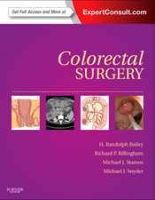 Colorectal Surgery: Expert Consult - Online and PrintH. Randolph Bailey-26%Preț: 872.64 lei1179.04 lei
 -
 Colon and Rectal Surgery: Anorectal OperationsSteven D. Wexner-23%Preț: 1530.21 lei1981.48 lei
 -
 Frontiers in Colorectal SurgeryRobin K S Phillips-19%Preț: 290.86 lei360.49 lei
 -
 -23%Preț: 1635.41 lei2122.59 lei
 -
 Current Common Dilemmas in Colorectal SurgeryChristopher M. Schlachta-5%Preț: 804.53 lei846.87 lei
 -
 CT Colonography Atlas: For the Practicing RadiologistEmanuele Neri-5%Preț: 704.77 lei741.87 lei
Public țintă
Professional/practitionerDe ce să citești această carte
Recomandăm această lucrare chirurgilor în formare și specialiștilor care doresc să își rafineze diagnosticul diferențial prin studii de caz reale. Cititorul câștigă o perspectivă integrată asupra patologiei „vii”, beneficiind de puncte cheie de predare (teaching points) extrase din cazuri unice. Este un instrument esențial pentru pregătirea examenelor de bord, oferind o corelație rară între anatomia chirurgicală și rezultatele histopatologice.
Despre autor
Dr. Mark Killingback este o figură proeminentă în chirurgia colorectală mondială, cu o carieră de peste 37 de ani dedicată acestui domeniu. Membru onorific al American College of Surgeons și al numeroaselor societăți chirurgicale europene, dr. Killingback este recunoscut pentru înființarea primei unități specializate de coloproctologie din Australia. Dincolo de expertiza clinică, autorul este apreciat pentru talentul său de a ilustra observațiile chirurgicale, transformând experiența acumulată la Sydney în materiale educaționale de înaltă precizie, menite să ghideze noile generații de chirurgi gastrointestinali.
Descriere scurtă
Visually, every chapter presents the reader with operative and/or diagnostic photos, and anatomic line drawings by the author. The text, more extensive than in many atlases, provides a concise yet complete operative record: patient history/work up, anatomic anomalies, the procedure itself, pathologic findings, and follow up.
Key teaching points emphasize the most important and unique aspects of every case. Residents, fellows, and even seasoned practitioners will gain valuable diagnostic and therapeutic insights from this material. The case study presentation provides an excellent review tool for the American Board of Colon and Rectal Surgeryexam.
Cuprins
Textul de pe ultima copertă
"The experienced surgeon will appreciate this book by recognizing the details and exquisitely rendered images that call to mind similar cases encountered. For the surgeon or trainee relatively new to the practice of colorectal surgery, the graphic presentation of the surgical pathology, with the accompanying succinct and informative text, will make the acquisition of this book a worthwhile one."
Victor Fazio, M.D. and Stanley Goldberg, M.D.
As Lord Moynihan frequently mentioned in his legendary lectures, surgeons have the unique opportunity to study pathology in vivo, or in a living state. The pathology is seen in relation to normal anatomy, leading to important surgical judgments that ensure only diseased tissue will be removed, and with minimal damage or sacrifice to normal structures. In addition to influencing surgical design making, observation of living pathology offers a glimpse into the disease process and providesa valuable assessment of the morphological features of the specimen before it is preserved with formalin.
Colorectal Surgery: Living Pathology in the Operating Room is a unique collection of beautifully illustrated cases focusing on the pathology as much as the operative technique used to remove the diseased tissue. One hundred case reports describe unusual findings, or unusual manifestations of more common conditions, encountered by the author during his 40-plus years in practice as a colorectal surgeon. All of the drawings in the book were sketched immediately after the procedures in order to capture the details and significance of the specimen. As finished drawings, Dr. Killingback’s beautiful images increase the reader’s conceptual knowledge of surgical pathology and how it presents. Accompanying text, photos, and diagnostic studies provide the details of patient management and shed further light on the intrinsic art and science of interpreting living pathology, making this a must-have resource for colorectal surgeons and residents as well as general surgeons and surgical pathologists.