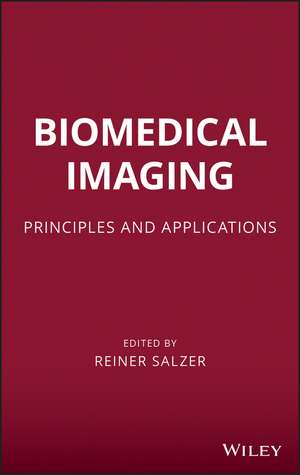
Biomedical Imaging
Editat de Reiner Salzeren Limba Engleză Hardback – 22 mai 2012
Preț: 929.67 lei
Preț vechi: 978.60 lei
-5%
Puncte Express: 1395
Carte tipărită la comandă
Livrare economică 18 noiembrie-02 decembrie
Livrare prin curier în România Termenul estimat este afișat lângă disponibilitate.
Transport gratuit pentru acest produs Plată online sau ramburs, în funcție de opțiunile comenzii.
Retur gratuit în 14 zile Comandă securizată și suport în română.
Specificații
ISBN-13: 9780470648476
ISBN-10: 0470648473
Pagini: 448
Dimensiuni: 161 x 240 x 28 mm
Greutate: 0.83 kg
Ediția:New.
Editura: Wiley
Locul publicării:Hoboken, United States
ISBN-10: 0470648473
Pagini: 448
Dimensiuni: 161 x 240 x 28 mm
Greutate: 0.83 kg
Ediția:New.
Editura: Wiley
Locul publicării:Hoboken, United States
V-ar putea interesa
-
 Human Cloning in the MediaJoan HaranPreț: 461.37 lei
 -
 CMOS BiomicrosystemsIniewski-9%Preț: 987.23 lei1084.87 lei
 -
 -15%Preț: 677.64 lei797.22 lei
 -
 Enhancing Extraction Processes in the Food IndustryNikolai Lebovka-15%Preț: 564.11 lei663.66 lei
 -
 Valorization of Food Processing By-ProductsM. Chandrasekaran-18%Preț: 823.11 lei1003.80 lei
 -
 Managing Biogas Plants: A Practical GuideMario Alejandro Rosato-18%Preț: 1260.06 lei1536.66 lei
 -
 -15%Preț: 422.09 lei496.58 lei
 -
 -14%Preț: 900.92 lei1047.58 lei
 -
 -9%Preț: 1197.86 lei1316.33 lei
 -
 Methionine Oxidation in Human Prion ProteinChristina Wolschner-8%Preț: 460.34 lei500.37 lei
 -
 NanoimagingBeth A. Goins-5%Preț: 1055.41 lei1110.95 lei
 -
 STEM CELL AND TISSUE ENGINEERINGLi Song-18%Preț: 961.84 lei1172.97 lei
Public țintă
Chemists, biologists and clinical researchers working at research institutes, universities, hospitals and pharmaceutical companies, e quipment developers, including optical and electronic engineers and chemical engineersNotă biografică
Reiner Salzer, PhD, is a professor at the Institute for Analytical Chemistry at Technische Universität in Dresden, Germany.
Cuprins
Preface xv
Contributors xvii
1 Evaluation of Spectroscopic Images 1
Patrick W.T. Krooshof, Geert J. Postma, Willem J. Melssen, and Lutgarde M.C. Buydens
1.1 Introduction 1
1.2 Data Analysis 2
1.2.1 Similarity Measures 3
1.3 Applications 11
References 25
2 Evaluation of Tomographic Data 30
Jorg van den Hoff
2.1 Introduction 30
2.2 Image Reconstruction 33
2.3 Image Data Representation: Pixel Size and Image Resolution 34
2.4 Consequences of Limited Spatial Resolution 39
2.5 Tomographic Data Evaluation: Tasks 46
2.6 Summary 61
References 61
3 X-Ray Imaging 63
Volker Hietschold
3.1 Basics 63
3.2 Instrumentation 66
3.3 Clinical Applications 76
3.4 Radiation Exposure to Patients and Employees 92
References 95
4 Computed Tomography 97
Stefan Ulzheimer and Thomas Flohr
4.1 Basics 97
4.2 Instrumentation 102
4.3 Measurement Techniques 109
4.4 Applications 119
4.5 Outlook 125
References 127
5 Magnetic Resonance Technology 131
Boguslaw Tomanek and Jonathan C. Sharp
5.1 Introduction 131
5.2 Magnetic Nuclei Spin in a Magnetic Field 133
5.3 Image Creation 139
5.4 Image Reconstruction 145
5.5 Image Resolution 148
5.6 Noise in the Image-SNR 149
5.7 Image Weighting and Pulse Sequence Parameters TE and TR 150
5.8 A Menagerie of Pulse Sequences 152
5.9 Enhanced Diagnostic Capabilities of MRI-Contrast Agents 159
5.10 Molecular MRI 159
5.11 Reading the Mind-Functional MRI 160
5.12 Magnetic Resonance Spectroscopy 161
5.13 MR Hardware 164
5.14 MRI Safety 171
5.15 Imaging Artefacts in MRI 173
5.16 Advanced MR Technology and Its Possible Future 175
References 175
6 Toward A 3D View of Cellular Architecture: Correlative Light Microscopy and Electron Tomography 180
Jack A. Valentijn, Linda F. van Driel, Karen A. Jansen, Karine M. Valentijn, and Abraham J. Koster
6.1 Introduction 180
6.2 Historical Perspective 181
6.3 Stains for CLEM 182
6.4 Probes for CLEM 183
6.5 CLEM Applications 193
6.6 Future Perspective 202
References 205
7 Tracer Imaging 215
Rainer Hinz
7.1 Introduction 215
7.2 Instrumentation 216
7.3 Measurement Techniques 228
7.4 Applications 234
References 244
8 Fluorescence Imaging 248
Nikolaos C. Deliolanis, Christian P. Schultz, and Vasilis Ntziachristos
8.1 Introduction 248
8.2 Contrast Mechanisms 249
8.3 Direct Methods: Fluorescent Probes 251
8.4 Indirect Methods: Fluorescent Proteins 252
8.5 Microscopy 253
8.6 Macroscopic Imaging/Tomography 260
8.7 Planar Imaging 260
8.8 Tomography 262
8.9 Conclusion 267
References 268
9 Infrared and Raman Spectroscopic Imaging 275
Gerald Steiner
9.1 Introduction 275
9.2 Instrumentation 278
9.3 Raman Imaging 282
9.4 Sampling Techniques 283
9.5 Data Analysis and Image Evaluation 285
9.6 Applications 292
References 301
10 Coherent Anti-Stokes Raman Scattering Microscopy 304
Annika Enejder, Christoph Heinrich, Christian Brackmann, Stefan Bernet, and Monika Ritsch-Marte
10.1 Basics 304
10.2 Theory 306
10.3 CARS Microscopy in Practice 309
10.4 Instrumentation 310
10.5 Laser Sources 311
10.6 Data Acquisition 314
10.7 Measurement Techniques 316
10.8 Applications 320
10.9 Conclusions 326
References 327
11 Biomedical Sonography 331
Georg Schmitz
11.1 Basic Principles 331
11.2 Instrumentation of Real-Time Ultrasound Imaging 341
11.3 Measurement Techniques of Real-Time Ultrasound Imaging 347
11.4 Application Examples of Biomedical Sonography 359
References 365
12 Acoustic Microscopy for Biomedical Applications 368
Jurgen Bereiter-Hahn
12.1 Sound Waves and Basics of Acoustic Microscopy 368
12.2 Types of Acoustic Microscopy 372
12.3 Biomedical Applications of Acoustic Microscopy 391
12.4 Examples of Tissue Investigations using SAM 403
12.4.1 Hard Tissues 404
12.4.2 Cardiovascular Tissues 405
12.4.3 Other Soft Tissues 406
References 406
Contributors xvii
1 Evaluation of Spectroscopic Images 1
Patrick W.T. Krooshof, Geert J. Postma, Willem J. Melssen, and Lutgarde M.C. Buydens
1.1 Introduction 1
1.2 Data Analysis 2
1.2.1 Similarity Measures 3
1.3 Applications 11
References 25
2 Evaluation of Tomographic Data 30
Jorg van den Hoff
2.1 Introduction 30
2.2 Image Reconstruction 33
2.3 Image Data Representation: Pixel Size and Image Resolution 34
2.4 Consequences of Limited Spatial Resolution 39
2.5 Tomographic Data Evaluation: Tasks 46
2.6 Summary 61
References 61
3 X-Ray Imaging 63
Volker Hietschold
3.1 Basics 63
3.2 Instrumentation 66
3.3 Clinical Applications 76
3.4 Radiation Exposure to Patients and Employees 92
References 95
4 Computed Tomography 97
Stefan Ulzheimer and Thomas Flohr
4.1 Basics 97
4.2 Instrumentation 102
4.3 Measurement Techniques 109
4.4 Applications 119
4.5 Outlook 125
References 127
5 Magnetic Resonance Technology 131
Boguslaw Tomanek and Jonathan C. Sharp
5.1 Introduction 131
5.2 Magnetic Nuclei Spin in a Magnetic Field 133
5.3 Image Creation 139
5.4 Image Reconstruction 145
5.5 Image Resolution 148
5.6 Noise in the Image-SNR 149
5.7 Image Weighting and Pulse Sequence Parameters TE and TR 150
5.8 A Menagerie of Pulse Sequences 152
5.9 Enhanced Diagnostic Capabilities of MRI-Contrast Agents 159
5.10 Molecular MRI 159
5.11 Reading the Mind-Functional MRI 160
5.12 Magnetic Resonance Spectroscopy 161
5.13 MR Hardware 164
5.14 MRI Safety 171
5.15 Imaging Artefacts in MRI 173
5.16 Advanced MR Technology and Its Possible Future 175
References 175
6 Toward A 3D View of Cellular Architecture: Correlative Light Microscopy and Electron Tomography 180
Jack A. Valentijn, Linda F. van Driel, Karen A. Jansen, Karine M. Valentijn, and Abraham J. Koster
6.1 Introduction 180
6.2 Historical Perspective 181
6.3 Stains for CLEM 182
6.4 Probes for CLEM 183
6.5 CLEM Applications 193
6.6 Future Perspective 202
References 205
7 Tracer Imaging 215
Rainer Hinz
7.1 Introduction 215
7.2 Instrumentation 216
7.3 Measurement Techniques 228
7.4 Applications 234
References 244
8 Fluorescence Imaging 248
Nikolaos C. Deliolanis, Christian P. Schultz, and Vasilis Ntziachristos
8.1 Introduction 248
8.2 Contrast Mechanisms 249
8.3 Direct Methods: Fluorescent Probes 251
8.4 Indirect Methods: Fluorescent Proteins 252
8.5 Microscopy 253
8.6 Macroscopic Imaging/Tomography 260
8.7 Planar Imaging 260
8.8 Tomography 262
8.9 Conclusion 267
References 268
9 Infrared and Raman Spectroscopic Imaging 275
Gerald Steiner
9.1 Introduction 275
9.2 Instrumentation 278
9.3 Raman Imaging 282
9.4 Sampling Techniques 283
9.5 Data Analysis and Image Evaluation 285
9.6 Applications 292
References 301
10 Coherent Anti-Stokes Raman Scattering Microscopy 304
Annika Enejder, Christoph Heinrich, Christian Brackmann, Stefan Bernet, and Monika Ritsch-Marte
10.1 Basics 304
10.2 Theory 306
10.3 CARS Microscopy in Practice 309
10.4 Instrumentation 310
10.5 Laser Sources 311
10.6 Data Acquisition 314
10.7 Measurement Techniques 316
10.8 Applications 320
10.9 Conclusions 326
References 327
11 Biomedical Sonography 331
Georg Schmitz
11.1 Basic Principles 331
11.2 Instrumentation of Real-Time Ultrasound Imaging 341
11.3 Measurement Techniques of Real-Time Ultrasound Imaging 347
11.4 Application Examples of Biomedical Sonography 359
References 365
12 Acoustic Microscopy for Biomedical Applications 368
Jurgen Bereiter-Hahn
12.1 Sound Waves and Basics of Acoustic Microscopy 368
12.2 Types of Acoustic Microscopy 372
12.3 Biomedical Applications of Acoustic Microscopy 391
12.4 Examples of Tissue Investigations using SAM 403
12.4.1 Hard Tissues 404
12.4.2 Cardiovascular Tissues 405
12.4.3 Other Soft Tissues 406
References 406
Descriere
This book presents and describes imaging technologies that can be used to study chemical processes and structural interactions in dynamic systems, principally in biomedical systems.