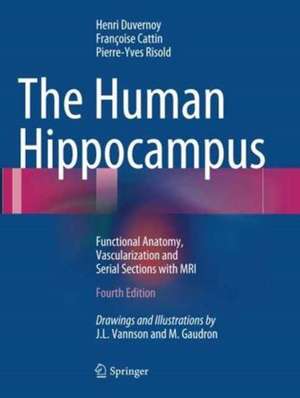
The Human Hippocampus
Autor Henri M. Duvernoy, Francoise Cattin, Pierre-Yves Risolden Limba Engleză Paperback – 3 iul 2013
Cea de-a patra ediție a lucrării The Human Hippocampus reprezintă un standard de precizie clinică în neuroanatomie, fiind actualizată pentru a reflecta progresele neurochirurgicale și tehnologiile avansate de imagistică medicală. Remarcăm în această revizuire integrarea sistematică a imaginilor RMN de înaltă rezoluție (3T), însoțite de desene explicative care clarifică raporturile anatomice complexe în planuri coronale, sagitale și axiale. Un element de noutate absolută față de edițiile precedente este includerea unor vederi RMN specifice pentru principalele afecțiuni hipocampale, transformând atlasul dintr-un instrument pur anatomic într-un suport diagnostic valoros. Structura volumului este riguros organizată, începând cu microanatomia structurilor fine — Cornu Ammonis și Gyrus Dentatus — și continuând cu o analiză detaliată a vascularizației. Descoperim aici o descriere minuțioasă a sistemului arterial și venos, atât la nivel superficial, cât și profund, esențială pentru siguranța intervențiilor în neurochirurgie. Autorul Henri M. Duvernoy, cunoscut pentru rigoarea demonstrată și în Human Brain Stem Vessels, menține aceeași abordare analitică, unde ilustrația nu este doar decorativă, ci servește drept ghid de orientare spațială. În comparație cu Nolte's The Human Brain in Photographs and Diagrams, care oferă o perspectivă generală asupra întregului sistem nervos central, lucrarea de față se concentrează exclusiv pe lobul temporal medial. Este o alternativă specializată la Atlas of Functional Neuroanatomy pentru rezidenții și specialiștii în neuroradiologie, având avantajul unei corelații directe între secțiunile anatomice reale și reprezentările RMN 3T. Deși The Hippocampus Book acoperă mai larg aspectele de fiziologie, volumul lui Duvernoy rămâne imbatabil în ceea ce privește cartografierea vasculară și anatomică necesară în practica chirurgicală.
Preț: 895.30 lei
Preț vechi: 942.42 lei
-5%
Carte disponibilă
Livrare economică 06-20 august
Livrare express 23-29 iulie pentru 47.33 lei
Specificații
ISBN-10: 3662495740
Pagini: 248
Ilustrații: VIII, 237 p. 133 illus., 13 illus. in color.
Dimensiuni: 210 x 279 x 13 mm
Greutate: 0.69 kg
Ediția:4th edition 2013
Editura: Springer
Locul publicării:Berlin, Heidelberg, Germany
V-ar putea interesa
-
 The Rhesus Monkey Brain in Stereotaxic CoordinatesGeorge Paxinos-31%Preț: 738.22 lei1064.62 lei
 -
 Structure of the Human Brain: A Photographic AtlasStephen J. DeArmond-5%Preț: 637.72 lei671.28 lei
 -
 Preț: 307.18 lei
 -
 Nolte's The Human Brain in Photographs and DiagramsTodd W. Vanderah-27%Preț: 335.66 lei460.28 lei
 -
 Imaging Anatomy Brain and SpineAnne G. Osborn-28%Preț: 1329.66 lei1846.90 lei
 -
 Netter's Head and Neck Anatomy for DentistryNeil S. Norton-25%Preț: 369.17 lei491.22 lei
 -
 Neuroanatomy: Illustrated Colour TextAlan R. Crossman-31%Preț: 262.29 lei379.52 lei
 -
 Netter's Neuroscience Coloring BookDavid L. Felten-25%Preț: 130.04 lei172.39 lei
 -
 Gray's Anatomy: The Anatomical Basis of Clinical PracticeSusan Standring-29%Preț: 1273.07 lei1783.78 lei
 -
 Imaging Anatomy: Head and NeckSurjith Vattoth-31%Preț: 1369.97 lei1984.11 lei
 -
 Principles of Cognitive NeuroscienceDale Purves-27%Preț: 1313.85 lei1799.80 lei
 -
 The Brain AtlasJoseph Hanaway-5%Preț: 470.07 lei494.82 lei
 -
 -22%Preț: 524.48 lei674.33 lei
 -
 Douglas Neuroanatomie. High-Yield™ NeuroanatomyDouglas J. Gould-26%Preț: 262.93 lei355.08 lei
 -
 Anatomic Basis of Neurologic DiagnosisCary Alberstone-5%Preț: 1206.74 lei1270.25 lei
 -
 -5%Preț: 671.59 lei706.94 lei
 -
 The Man Who Folded HimselfDavid Gerrold-44%Preț: 66.52 lei118.22 lei
 -
 The NeuronomiconGrayson W. Hooper-5%Preț: 434.27 lei457.12 lei
 -
 Pocket Atlas of Sectional Anatomy, Volume I: Head and NeckTorsten Bert Möller-5%Preț: 367.32 lei386.66 lei
 -
 The Amygdaloid Nuclear ComplexVincent Di Marino-5%Preț: 702.46 lei739.44 lei
De ce să citești această carte
Această ediție este esențială pentru neurochirurgi și neuroradiologi care au nevoie de o hartă anatomică de mare precizie a hipocampului. Cititorul câștigă capacitatea de a interpreta corect imaginile RMN 3T prin corelarea lor cu secțiunile macroscopice detaliate. Este un instrument de lucru clinic care elimină ambiguitățile în localizarea structurilor fine și a rețelelor vasculare, crucial în planificarea preoperatorie și în diagnosticul neurologic de finețe.
Despre autor
Henri M. Duvernoy este un anatomist de renume internațional, a cărui carieră a fost dedicată cartografierii detaliate a creierului uman. Lucrările sale sunt publicate constant de Springer și sunt recunoscute pentru acuratețea diagramelor și a secțiunilor anatomice. Expertiza sa în angioarhitectonica cerebrală este evidențiată în volume de referință precum cele despre vasele trunchiului cerebral sau venele superficiale ale creierului. Abordarea sa interdisciplinară îmbină anatomia clasică cu cele mai noi tehnici de imagistică prin rezonanță magnetică, oferind comunității medicale instrumente fundamentale pentru neurochirurgia modernă.
Descriere scurtă
Cuprins
Recenzii
“This is a comprehensive atlas and correlated functional anatomy text of CA1-CA3, body, head, and the circuitry along with the vascular supply to this area of the temporal lobe and surrounding structures. … The book will be a good addition to the shelves of neuroscientists, epileptologists, neurosurgeons, neurology professionals, and students for a quick comprehensive look at the complexity of the human hippocampus.” (Joseph J. Grenier, Amazon.com, March, 2015)
Notă biografică
Françoise Cattin completed her medical thesis in 1980 and since 1986 has worked at CHU Besançon, France. In 1996-7 she completed a Visiting Professorship at Montreal Neurological Hospital, McGill University, Canada. Her memberships include the Société Française de Neuroradiologie, the SociétéFrançaise de Radiologie, and the Société européenne de Neuroradiologie. Dr. Cattin is the author of a number of articles in peer-reviewed journals and has co-authored or co-edited several previous books, including Computed Tomography of the Pituitary Gland (Springer, 1986) and three editions of Echo-Doppler des artères carotides et vertébrales: aspects pratiques (Masson).
Pierre-Yves Risold completed his PhD thesis at the Histology Laboratory, Faculté de Médecine et de Pharmacie, Université de Franche-Comté, Besançon in 1991. He subsequently performed postdoctoral studies at the University of Southern California, Los Angeles, in the course of which he analyzed the anatomical relationships of the hippocampus with the septum and hypothalamus. As CR1 INSERM he returned to the Histology Laboratory (EA 3922) in Besançon in 1997. Dr. Risold is a member of the Société de Neuroendocrinologie and Société des Neurosciences. He has published more than 50 articles in peer-reviewed journals as well as several book chapters.