The Eye Anatomical Chart
Autor Anatomical Chart Companyen Limba Engleză Wallchart – 25 apr 2025
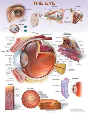
- Outer eye with surface anatomy, anterior view, also showing the lacrimal gland and nasolacrimal duct
- Eyeball in skull, lateral and top views
- Visual field diagram
- Cross section of the eye, lateral view, as the large central illustration along with the anterior chamber angle
- Medial cross section of the eye
- The cornea, macula lutea, fundus, and retina in close-up views
Consultant (2025): Christopher J. Rapuano, MD, Wills Eye Hospital
Original medical illustrations by Keith Kasnot, CMI, in consultation with Randall Paul, OD, Phoenix, Arizona
Preț: 93.62 lei
Preț vechi: 98.55 lei
-5%
Puncte Express: 140
Disponibil
Livrare economică 22 mai-03 iunie
Specificații
ISBN-13: 9781975255411
ISBN-10: 1975255410
Pagini: 1
Dimensiuni: 508 x 660 mm
Greutate: 0.1 kg
Ediția:Second
Editura: LWW
Colecția Lww
ISBN-10: 1975255410
Pagini: 1
Dimensiuni: 508 x 660 mm
Greutate: 0.1 kg
Ediția:Second
Editura: LWW
Colecția Lww
V-ar putea interesa
-
 -34%Preț: 470.54 lei714.50 lei
 -
 Clinical Anatomy and Physiology of the Visual SystemLee Ann Remington-30%Preț: 539.19 lei769.29 lei
 -
 Netter's Head and Neck Anatomy for DentistryNeil S. Norton-33%Preț: 319.91 lei475.58 lei
 -
 Adler's Physiology of the EyeLeonard A. Levin-5%Preț: 667.70 lei702.84 lei
 -
 Ocular PathologyMyron Yanoff-24%Preț: 1891.78 lei2499.40 lei
 -
 Video Atlas of Ophthalmic UltrasoundBernadete Ayres-30%Preț: 963.88 lei1371.03 lei
 -
 Kanski's Clinical Ophthalmology: A Systematic ApproachJohn F. Salmon-28%Preț: 1167.86 lei1629.18 lei
 -
 An Atlas of Animal Anatomy for ArtistsW. Ellenberger-19%Preț: 95.06 lei117.76 lei
 -
 -5%Preț: 532.60 lei560.63 lei
 -
 Kanski's Synopsis of Clinical OphthalmologyJohn F. Salmon-29%Preț: 476.81 lei670.97 lei
 -
 -25%Preț: 972.09 lei1303.98 lei
 -
 Anatomy of the Heart Anatomical ChartAnatomical Chart Company-5%Preț: 129.35 lei136.15 lei
 -
 -5%Preț: 134.13 lei141.19 lei
 -
 -5%Preț: 129.67 lei136.50 lei
 -
 -5%Preț: 703.43 lei740.45 lei
 -
 -37%Preț: 123.99 lei198.15 lei
 -
 The Digestive System Anatomical ChartAnatomical Chart Company-5%Preț: 133.32 lei140.34 lei
 -
 Ear, Nose, and Throat Anatomical ChartAnatomical Chart Company-5%Preț: 89.10 lei93.78 lei
 -
 Disorders of the Eye Anatomical ChartAnatomical Chart Company-5%Preț: 93.41 lei98.32 lei
Descriere
The Eye Anatomical Chart, Second Edition features general anatomy of the eye with colorful detailed illustrations, all fully labeled.
Consultant (2025): Christopher J. Rapuano, MD, Wills Eye Hospital
Original medical illustrations by Keith Kasnot, CMI, in consultation with Randall Paul, OD, Phoenix, Arizona
- Outer eye with surface anatomy, anterior view, also showing the lacrimal gland and nasolacrimal duct
- Eyeball in skull, lateral and top views
- Visual field diagram
- Cross section of the eye, lateral view, as the large central illustration along with the anterior chamber angle
- Medial cross section of the eye
- The cornea, macula lutea, fundus, and retina in close-up views
Consultant (2025): Christopher J. Rapuano, MD, Wills Eye Hospital
Original medical illustrations by Keith Kasnot, CMI, in consultation with Randall Paul, OD, Phoenix, Arizona