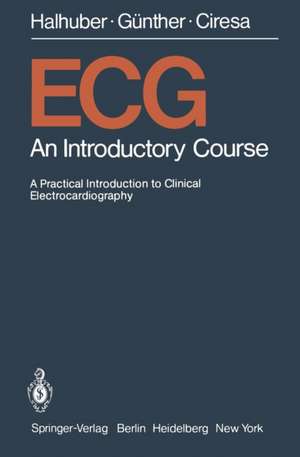
ECG
Autor M. J. Halhuber, R. Günther, M. Ciresa Traducere de H. J. Hirschen Limba Engleză Paperback – iul 1979
Preț: 347.69 lei
Preț vechi: 365.99 lei
-5%
Puncte Express: 522
Carte tipărită la comandă
Livrare economică 30 noiembrie-14 decembrie
Livrare prin curier în România Termenul estimat este afișat lângă disponibilitate.
Transport gratuit de la 400.00 lei Plată online sau ramburs, în funcție de opțiunile comenzii.
Retur gratuit în 14 zile Comandă securizată și suport în română.
Specificații
ISBN-13: 9783540093268
ISBN-10: 3540093265
Pagini: 172
Ilustrații: XII, 158 p. 6 illus.
Dimensiuni: 133 x 203 x 10 mm
Greutate: 0.2 kg
Editura: Springer
Locul publicării:Berlin, Heidelberg, Germany
ISBN-10: 3540093265
Pagini: 172
Ilustrații: XII, 158 p. 6 illus.
Dimensiuni: 133 x 203 x 10 mm
Greutate: 0.2 kg
Editura: Springer
Locul publicării:Berlin, Heidelberg, Germany
V-ar putea interesa
-
 Cardiac Drug TherapyM. Gabriel Khan-5%Preț: 1187.52 lei1250.03 lei
 -
 Introduction to Translational Cardiovascular ResearchDennis V. Cokkinos-5%Preț: 1172.12 lei1233.81 lei
 -
 Clinico-Pathological Atlas of Cardiovascular DiseasesJoaquín S. Lucena-5%Preț: 1269.24 lei1336.04 lei
 -
 Drowning: Prevention, Rescue, TreatmentJoost J.L.M. Bierens-5%Preț: 1604.08 lei1688.51 lei
 -
 Congenital Heart DiseaseHideaki Senzaki-5%Preț: 737.00 lei775.79 lei
 -
 Coronary Microvascular DysfunctionFilippo Crea-5%Preț: 724.75 lei762.89 lei
 -
 Text Atlas of Practical ElectrocardiographyMassimo Romanò-5%Preț: 633.37 lei666.70 lei
 -
 Sudden DeathH.E. Kulbertus-5%Preț: 364.56 lei383.75 lei
Public țintă
ResearchCuprins
1. Vectorcardiography.- 2. Usual ECG Leads and Their Interrelation.- 3. Interpretation of the “Electric” Axis of the Heart.- 4. The Normal ECG.- 4.1 The P Wave.- 4.2 The AV Interval (PQ or PR).- 4.3 The QRS Complex.- 4.3.1 QRS Amplitude.- 4.3.2 QRS Duration.- 4.3.3 QR Interval (Intrinsicoid Deflection).- 4.4 The ST-T Segment.- 4.5 The T Wave.- 4.6 The QT Interval.- 4.7 The U Wave.- 5. Memory Aid to Systematic ECG Description and Evaluation.- 6. Ventricular Conduction Disorder — Bundle Branch Block.- 6.1 Unifascicular Block.- 6.2 Bifascicular Bundle Branch Block.- 6.3 Trifascicular Blocks.- 7. The WPW Syndrome.- 8. ECG in Hypertrophy of Individual Chambers of the Heart.- 8.1 ECG in Hypertrophy of the Atria.- 8.2 ECG in Hypertrophy of the Ventricles.- 8.2.1 Depolarization.- 8.2.2 Repolarization.- 8.2.3 Position of the Cardiac Axis.- 9. ECG in Myocardial Infarction.- 9.1 Hypotheses of Necrosis, Injury, Ischemia.- 9.2 Sites of Myocardial Infarction.- 9.3 Course and Classification of Infarcts.- 9.4 Differential Diagnosis of the ECG in Myocardial Infarction.- 9.5 Infarct and Bundle Branch Block.- 10. Changes in the ST-T Segment.- 10.1 Causes of ST-T Changes.- 11. Exercise ECG.- 11.1 Changes in the Exercise ECG Suggestive of Coronary Disease.- 11.2 Doubtful and Prognostically Unreliable Changes in the Exercise ECG.- 11.3 Absolute Contraindications to the Exercise Tolerance Test or Bicycle Dynamometry.- 11.4 Relative Contraindications.- 11.5 Criteria for Discontinuance.- 12. ECG Diagnosis of Arrhythmias.- 12.1 Are (normal) P Waves Present?.- 12.2 What is the Distance Between Individual P Waves?.- 12.3 What is the Shape of the P waves?.- 12.4 What is the Distance of P Waves from the Following QRS Complex?.- 12.5 What is the Distance Between Apparently Identical VentricularComplexes?.- 12.6 What is the Time Relation of Differently Shaped Ventricular Complexes to Each Other or to the Basic Rhythm?.- 12.7 Incidence of Different Types of Arrhythmia.- 13. Pacemaker ECG.- 13.1 Fixed-Rate Pacemakers.- 13.2 Demand Pacemakers.- 13.3 Atrial-Triggered Pacemakers.- 13.4 Bifocal Demand Pacemakers.- 13.5 Complications After Pacemaker Implantation.- 14. ECG in Children.- 14.1 Normal Development of ECG as a Whole.- 14.2 Characteristic Patterns of Childhood Tracings.- 14.3 Effects of Extracardiac Factors.- 14.4 Technical Difficulties.- 15. Technique of ECG Recording.- 16. Concluding Cautionary Remarks on ECG Interpretation.- 16.1 Cardiac Arrhythmias.- 16.2 Diagnosis of Infarction.- 16.3 Suspected Hypertrophy of Individual Parts of the Heart.