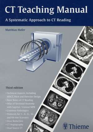
CT Teaching Manual: A Systematic Approach to CT Reading
Autor Matthias Hoferen Limba Engleză Paperback – 21 aug 2007
Bazându-ne pe protocoalele actuale de imagistică și pe cerințele tehnice ale radiologiei moderne, CT Teaching Manual se impune ca o resursă clinică fundamentală pentru formarea radiologilor și a tehnicienilor. Observăm că ediția a cincea integrează progresele tehnologice recente, precum scanarea 64-slice și sistemele dual-source CT, elemente esențiale pentru obținerea unor reconstituiri MPR (coronale și sagitale) de înaltă precizie. Structura manualului urmărește o progresie logică: începe cu bazele fizice și tehnice, trece prin regulile fundamentale de citire a examinării și administrarea substanțelor de contrast, culminând cu secțiuni detaliate de patologie pe regiuni anatomice. Remarcăm echilibrul vizual susținut de cele 1243 de figuri; acolo unde Computed Tomography for Technologists pune accent pe pregătirea pentru examenele de certificare și îngrijirea pacientului, volumul semnat de Matthias Hofer aprofundează corelația directă dintre desenele anatomice explicative și imaginile CT patologice. Această abordare este o semnătură a autorului, pe care o regăsim și în Ultrasound Teaching Manual, unde procesul de învățare este segmentat în pași sistematici. Recomandăm acest manual pentru rigoarea cu care tratează dozimetria și siguranța radiațiilor, dar și pentru includerea unor module de nișă, cum ar fi introducerea în PET/CT și angiografia CT, oferind o perspectivă clinică integrată asupra diagnosticului imagistic modern.
Preț: 414.91 lei
Preț vechi: 436.74 lei
-5%
Carte indisponibilă temporar
Specificații
ISBN-10: 1588905810
Pagini: 224
Ilustrații: 650
Dimensiuni: 210 x 292 x 13 mm
Greutate: 0.89 kg
Ediția:3rd edition
Editura: Thieme
Colecția Thieme
V-ar putea interesa
-
 The Vatican Israel AccordsMarshall BregerPreț: 445.73 lei
 -
 The Key to the Brescia CasketCatherine Brown Tkacz-23%Preț: 592.45 lei769.42 lei
 -
 The Intellectual Appeal of Catholicism and the Idea of a Catholic UniversityMark William Roche-23%Preț: 581.28 lei754.92 lei
 -
 Racism in MindMichael P. Levine-27%Preț: 771.72 lei1057.15 lei
 -
 Unwelcome and Unlawful – Sexual Harassment in the American WorkplaceRaymond F. Gregory-27%Preț: 770.24 lei1055.12 lei
 -
 -27%Preț: 768.75 lei1053.09 lei
 -
 In the Shadow of "Just Wars" – Violence, Politics and Humanitarian ActionMédecins Sans F Médecins Sans F-27%Preț: 771.45 lei1056.79 lei
 -
 -23%Preț: 727.52 lei944.83 lei
 -
 In Search of the Rain ForestCandace SlaterPreț: 251.84 lei
 -
 Gender and National LiteratureTomiko YodaPreț: 249.71 lei
 -
 Living Spirit, Living PracticeRuth FrankenbergPreț: 249.16 lei
 -
 Jorge Semprún – The Spaniard Who Survived the Nazis and Conquered ParisSoledad Fox Maura-27%Preț: 850.02 lei1164.40 lei
 -
 Teaching Manual of Color Duplex SonographyMatthias Hofer-5%Preț: 566.74 lei596.57 lei
 -
 The Chest X–Ray – A Systematic Teaching AtlasMatthias HoferPreț: 272.67 lei
 -
 -5%Preț: 661.57 lei696.39 lei
 -
 Preț: 163.55 lei
 -
 MuralsE MinguetPreț: 147.23 lei
De ce să citești această carte
Recomandăm această lucrare oricărui rezident în radiologie sau tehnician care dorește să treacă de la teoria de bază la interpretarea clinică precisă. Cititorul câștigă o metodologie structurată de analiză a imaginilor și acces la peste 1200 de ilustrații de înaltă calitate. Este un instrument practic de învățare care, prin chestionarele de autoevaluare, garantează stăpânirea protocoalelor de scanare și a anatomiei secționale.
Despre autor
Matthias Hofer este un autor recunoscut în literatura medicală de specialitate, cunoscut pentru capacitatea sa de a transforma subiecte complexe de imagistică în ghiduri didactice accesibile. Opera sa include titluri de referință precum Teaching Manual of Color Duplex Sonography și The Chest X–Ray – A Systematic Teaching Atlas, lucrări ce reflectă un interes constant pentru instruirea sistematică a personalului medical. Stilul său se caracterizează prin utilizarea extensivă a materialului vizual și a structurilor de tip curs, fiind adaptat nevoilor practice ale studenților și medicilor aflați în primii ani de specializare în radiologie și ecografie.
Descriere scurtă
Recenzii
Notă biografică
Cuprins
Basic Rules for Reading CT Examinations
Preparing the Patient
Administration of Contrast Agents
Cranial CT
Cranial CT: Normal Findings
Cranial Pathology
Cervical CT
Cervical Pathology
Chest CT
Chest CT Pathology
-Chest Wall
-Mediastinum
-Lung
Abdominal CT
Abdominal Pathology
-Abdominal Wall
-Liver
-Biliary Tract
-Gallbladder
-Spleen
-Pancreas
-Adrenal Glands
-Kidneys
-Urinary Bladder
-Reproductive Organs
-Gastrointestinal Tract
-Retroperitoneum
-Skeletal Changes
Spinal Column: Skeletal Pathology
Lower Extremity
Radiation Safety
CT Angiography
Contrast Injectors
Dual Source CT
Introduction to PET/CT
Anatomy in Coronal MPRs
Anatomy in Sagittal MPRs