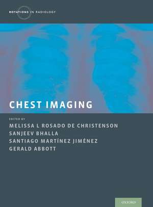
Chest Imaging: Rotations in Radiology
Editat de Melissa Rosado de Christenson, Sanjeev Bhalla, Gerald Abbott, Santiago Martinez-Jimenezen Limba Engleză Hardback – 25 oct 2019


Preț: 1219.46 lei
Preț vechi: 1629.54 lei
-25%
Puncte Express: 1829
Carte disponibilă
Livrare economică 06-17 noiembrie
Livrare prin curier în România Termenul estimat este afișat lângă disponibilitate.
Transport gratuit pentru acest produs Plată online sau ramburs, în funcție de opțiunile comenzii.
Retur gratuit în 14 zile Comandă securizată și suport în română.
Specificații
ISBN-13: 9780199858064
ISBN-10: 0199858063
Pagini: 600
Dimensiuni: 223 x 282 x 32 mm
Greutate: 2.06 kg
Editura: Oxford University Press
Colecția OUP USA
Seria Rotations in Radiology
Locul publicării:New York, United States
ISBN-10: 0199858063
Pagini: 600
Dimensiuni: 223 x 282 x 32 mm
Greutate: 2.06 kg
Editura: Oxford University Press
Colecția OUP USA
Seria Rotations in Radiology
Locul publicării:New York, United States
V-ar putea interesa
-
 Thoracic ImagingSujal R. Desai-22%Preț: 607.00 lei777.02 lei
 -
 Problem Solving in Chest ImagingSubba R. Digumarthy-28%Preț: 799.44 lei1104.54 lei
 -
 Thoracic Imaging The RequisitesJo-Anne O Shepard-27%Preț: 595.07 lei819.20 lei
 -
 Chest Radiology: Patterns and Differential DiagnosesJames C. Reed-28%Preț: 641.45 lei890.12 lei
 -
 ExpertDDx: ChestBrett W. Carter-28%Preț: 1387.10 lei1925.53 lei
 -
 Diagnostic Imaging: ChestMelissa L. Rosado-de-Christenson-28%Preț: 1884.68 lei2617.52 lei
 -
 Learning Radiology: Recognizing the BasicsWilliam Herring-28%Preț: 318.56 lei440.03 lei
 -
 Anatomy for Diagnostic ImagingStephanie Ryan-28%Preț: 424.24 lei592.90 lei
 -
 Imaging Anatomy: Chest, Abdomen, PelvisSiva P. Raman-5%Preț: 1847.60 lei1944.84 lei
 -
 Specialty Imaging: HRCT of the LungSantiago Martínez-Jiménez-28%Preț: 1453.50 lei2014.17 leiRecomandat
 -
 The Chest X-Ray: A Survival GuideGerald de Lacey-27%Preț: 322.43 lei444.20 lei
 -
 Rapid Review of RadiologyShahid Hussain-5%Preț: 784.34 lei825.62 lei
 -
 Imaging of the Chest, 2-Volume Set: Expert Radiology SeriesNestor L. Muller-30%Preț: 1502.47 lei2140.52 lei
 -
 Thoracic Imaging: A Core ReviewStephen Hobbs MD-5%Preț: 584.64 lei615.41 lei
 -
 Differential Diagnosis in Computed TomographyFrancis A. Burgener-5%Preț: 1726.26 lei1817.11 lei
 -
 The Chest X–Ray – A Systematic Teaching AtlasMatthias HoferPreț: 272.67 lei
 -
 Emergency Radiology of the Chest and Cardiovascular SystemMariano Scaglione-5%Preț: 1353.55 lei1424.78 lei
 -
 Diagnostic Imaging for Thoracic SurgeryMichele Anzidei-5%Preț: 1057.92 lei1113.60 lei
 -
 Imaging for Pediatricians: 100 Key CasesMaría I. Martínez-León-5%Preț: 362.27 lei381.34 lei
 -
 Diseases of the Chest and HeartJürg Hodler-5%Preț: 374.56 lei394.27 lei
Notă biografică
Melissa L Rosado-de-Christenson is a Professor of Clinical Radiology and the Section Chief for Thoracic Radiology at the University of Missouri-Kansas City, St. Luke's Hospital in Kansas City, Missouri.Sanjeev Bhalla is an Associate Professor of Radiology at Washington University and the Chief of Cardiothoracic Imaging at the Mallinckrodt Institute of Radiology in St. Louis, Missouri.Gerald Abbott is an Associate Radiologist in the Division of Thoracic Imaging at Massachusetts General Hospital and Harvard Medical School in Boston, Massachusetts.Santiago Martinez-Jiminez is an Assistant Professor of Clinical Radiology at the University of Missouri-Kansas City, St. Luke's Hospital in Kansas City, Missouri.
Cuprins
- Part I. Introduction to Chest Radiology
- 1. Introduction to Chest Radiology
- 2. Imaging Modalities
- 3. Overview of Normal Thoracic Imaging Anatomy
- 4. The radiology report and radiologic interpretation
- 5. Basic signs in chest radiology
- Part II Bedside Radiography
- 6. Introduction to Portable Chest Radiography: Support Devices
- 7. Endotracheal and Enteric Tubes
- 8. Vascular Catheters
- 9. Pacers / AICD
- 10. Thoracostomy tubes, mediastinal drains
- Part III Bedside Radiography - Common abnormalities
- 11 Bedside Chest Radiography-Common Findings
- 12. Pulmonary venous hypertension and edema
- 13. Airspace Disease - Atelectasis, pneumonia and aspiration
- 14. Acute respiratory distress syndrome
- Part IV Volume Loss
- 15. Volume Loss
- 16. Atelectasis: Opaque hemithorax
- 17. Upper and middle lobe atelectasis
- 18. Lower lobe atelectasis
- 19. Subsegmental and rounded atelectasis
- Part V Emergency Radiology
- 20. Introduction to Emergency Chest Radiology
- 21. Traumatic vascular injury
- 22. Chest trauma; Contusions, lacerations, fractures
- 23. Acute aortic syndromes and aortitis
- 24. Pulmonary thromboembolic disease
- 25. Sickle cell disease
- 26. Pulmonary hypertension
- Part VI Pleural disease
- 27. Introduction to Pleural disease
- 28. Pneumothorax
- 29. Effusion
- 30. Empyema and complications
- 31. Pleural thickening and calcification
- 32. Pleural neoplasms
- Part VII Pulmonary infections
- 33. Pulmonary infections
- 34. Community acquired pneumonia
- 35. Cavitary nodules, masses and consolidations
- 36. Endemic fungal pneumonias
- 37. Opportunistic fungal pneumonias
- 38. Tuberculosis
- 39. Non tuberculous mycobacterial infection
- 40. Mycoplasma and Viral pneumonias
- Part VIII The immunocompromised patient
- 41. The immunocompromised patient
- 42. The immunocompromised patient - AIDS
- 43. The immunocompromised patient - Non-AIDS
- Part IX Neoplasms of the lung and tracheobronchial tree
- 44. Introduction to neoplasms of lung and tracheobronchial tree
- 45. Lung cancer
- 46. Nodules and masses
- 47. Atelectasis and post-obstructive pneumonia
- 48. Lymphadenopathy and extrapulmonary involvement
- 49. Lung Cancer Staging
- 50. Bronchial carcinoid and bronchial gland carcinomas
- 51. Pulmonary metastases
- 52. Hamartoma and benign tumor-like lesions
- Part X Airways Disease
- 53. Airways Disease
- 54. Tracheal narrowing and tracheomalacia
- 55. Bronchiectasis
- 56. Emphysema
- 57. Bronchiolitis
- Part XI Connective Tissue Disorders and Autoimmune Conditions
- 58. Introduction to Connective Tissue Disorders and Autoimmune Conditions
- 59. Rheumatoid arthritis
- 60. Systemic Sclerosis
- 61. Pulmonary Vasculitis
- 62. Other autoimmune disorders
- 63. Eosinophilic lung diseases
- 64. Amyloidosis
- Part XII Pneumoconiosis
- 65. Pneumoconiosis
- 66. Asbestosis
- 67. Silocosis and Coal Worker's Pneumoconiosis
- Part XIII Iatrogenic conditions
- 68. Introduction to Iatrogenic Conditions
- 69. Radiation pneumonitis and fibrosis
- 70. Drug-induced lung disease
- 71. Post-operative complications
- Part XIV Diffuse Infiltrative Lung Disease
- 72. Diffuse Infiltrative Lung Disease
- 73. Ground glass opacity
- 74. Cystic lung disease
- 75. Pulmonary micronodules
- 76. Sarcoidosis
- Part XV Idiopathic interstitial pneumonias
- 77. Idiopathic interstitial pneumonias
- 78. Idiopathic pulmonary fibrosis
- 79. Non-Specific interstitial pneumonia
- 80. Cryptogenic organizing pneumonia
- Part XVI Mediastinal Abnormalities
- 81. Introduction to Mediastinal Abnormalities
- 82. Primary neoplasms
- 83. Lymphadenopathy
- 84. Cysts
- 85. Glandular enlargement
- 86. Vascular lesions including lymphatic malformations and aneurysms
- 87. Miscellaneous, including herniations and EMH
- Part XVII Developmental Abnormalities
- 88. Introduction to developmental abnormalities
- 89. Congenital bronchial atresia
- 90. Sequestration
- 91. Abnormal aorta and aortic branching
- 92. Abnormalities of pulmonary venous return
- 93. Abnornalities of systemic veins
- 94. Shunts including intracardiac and intrapulmonary
- Part XVIII Diseases of the Chest Wall and Diaphragm
- 95. Diseases of the Chest Wall and Diaphragm
- 96. Chest wall abnormalities
- 97. Diaphragmatic abnormalities