Broken Bones
Autor Felix S Chew, Catherine Maldijan, Hyojeong Mulcahyen Limba Engleză Paperback – 3 mai 2016
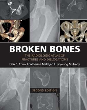
Remarcăm în această a doua ediție a lucrării Broken Bones o abordare riguros structurată a traumatologiei, care debutează cu analiza detaliată a fracturilor și luxațiilor degetelor, de la falangele distale până la articulațiile interfalangiene. Autorii, conduși de Felix S Chew, utilizează un format bazat pe dovezi vizuale masive, integrând 434 de cazuri clinice care acoperă întreg spectrul scheletic. Suntem de părere că progresia logică a capitolelor — de la extremitățile superioare către coloana cervicală, toraco-lombară și, ulterior, către membrul inferior — oferă o hartă diagnostică indispensabilă în mediul clinic. Fiecare entitate patologică este susținută de peste 1.100 de imagini radiologice ce ilustrează nu doar prezentările clasice, ci și variantele atipice care pot induce erori de diagnostic. Un element distinctiv al acestui volum este includerea unor secțiuni specializate pentru radiologia pediatrică și, în mod particular, pentru traumatismele complexe provocate de proiectile sau suflul exploziilor non-militare. Din punct de vedere stilistic, textul menține un ton precis și concis, punând accent pe corelația directă dintre imaginea radiografică și mecanismul de producere a leziunii. În contextul literaturii de specialitate, Broken Bones completează Skeleton Atlas prin adăugarea cazuisticii reale și a dinamicii clinice peste bazele anatomice. De asemenea, lucrarea se distinge de Handbook of Fractures prin focalizarea sa pe interpretarea imagistică extensivă, oferind o bibliotecă vizuală mult mai bogată pentru medicul radiolog sau ortoped aflat în fața unui caz incert.
Preț: 711.08 lei
Preț vechi: 748.51 lei
-5%
Carte disponibilă
Livrare economică 18 iunie-02 iulie
Specificații
ISBN-10: 1107499232
Pagini: 406
Ilustrații: 110 b/w illus.
Dimensiuni: 220 x 279 x 18 mm
Greutate: 1.14 kg
Ediția:2nd edition
Editura: Cambridge University Press
Locul publicării:New York, United States
V-ar putea interesa
-
 Emergency Medicine Board ReviewDanielle Campagne-22%Preț: 863.37 lei1108.86 lei
 -
 Tales of the Teahouse RetoldKatherine Liang ChewPreț: 143.47 lei
 -
 Emergency Management of Infectious DiseasesRachel L Chin-5%Preț: 1075.28 lei1131.87 lei
 -
 Introduction to Basic Cardiac DysrhythmiasSandra Atwood-5%Preț: 542.49 lei571.04 lei
 -
 Sander's Paramedic 5e W/Advantage AccessSanders, Mick-7%Preț: 3422.27 lei3687.96 lei
 -
 Arrhythmia Recognition: the Art of InterpretationTomas B. Garcia-54%Preț: 594.43 lei1280.53 lei
 -
 -23%Preț: 770.97 lei995.56 lei
 -
 The Washington Manual of Emergency MedicineMark D Levine MD, FACEP-22%Preț: 471.95 lei607.76 lei
 -
 First Responder Care EssentialsKris Lethbridge-5%Preț: 280.13 lei294.87 lei
 -
 Ambulance Care PracticeRichard Pilbery-5%Preț: 424.63 lei446.97 lei
De ce să citești această carte
Recomandăm această lucrare medicilor rezidenți și specialiștilor din radiologie și ortopedie care au nevoie de o referință vizuală rapidă. Cititorul câștigă acces la o bază de date vastă de peste 1.100 de imagini, învățând să identifice chiar și cele mai subtile fracturi. Este un instrument esențial pentru rafinarea diagnosticului diferențial în urgențele traumatologice.
Despre autor
Felix S Chew este un renumit specialist în radiologie musculo-scheletică, fiind recunoscut pentru contribuțiile sale academice și clinice în interpretarea imaginilor traumatice. Alături de Catherine Maldijan și Hyojeong Mulcahy, acesta a consolidat o metodologie de predare bazată pe cazuri clinice reale, menită să pună în punte teoria anatomică cu practica din departamentele de urgență. Expertiza autorilor se reflectă în selecția riguroasă a imaginilor și în claritatea explicațiilor ce însoțesc fiecare tip de fractură prezentat în acest volum publicat de Cambridge University Press.